All published articles of this journal are available on ScienceDirect.

Assessing the Structure of Tear Ferning in a Group of Healthy Young Subjects: A Pilot Study
Abstract
Purpose:
The purpose of the current pilot study is to investigate the structure of tear ferning in female and male subjects of different ages.
Methods:
A total of six normal subjects participated in the current pilot cross-sectional study. The female subjects were 11, 28, and 42 years old, whereas the male subjects were 28, 32, and 34 years old. Five tear samples were collected from each subject using capillary tubes. A small drop (1 μl) of tears was dried on a glass slide and observed under a light microscope (Olympus BX1). Rolando (four grades) and Masmali (five grades) scales were used to classify the tear ferning patterns.
Results:
The tear film pattern of all the subjects was surrounded by three thick and homogenous peripheral layers. The layers surrounding the ferning were better distinguished in the 11-year-old female compared to the layers around the ferning in other subjects. The tear ferning of the 11-year-old female was very fine without any spacing and considered as a Grade 0 (Masmali), Type 1 (Roland), whereas the 28-year-old female had Grade 3 (Masmali), Type 4 (Rolando). The 28-year-old male had Grade 1 (Masmali), Type 2 (Rolando), whereas the 34-year-old male and 42-year-old male had ferning of Grade 3 (Masmali), Type 4 (Rolando).
Conclusion:
The current pilot study showed that irrespective of gender, ferning patterning and grading degenerated and increased, respectively, as the age of the participants increased. Furthermore, the grading of ferning was better in male participants compared to female participants.
1. INTRODUCTION
The cornea is the outermost transparent surface of the eye which is covered anteriorly by tear fluid. The tear fluid provides hydration, lubrication, metabolite removal, and nourishment to the corneal epithelium and keeps the corneal surface smooth to maintain transparency [1]. The protection, stability and nutrition provided to the cornea by the tear film are due to the presence of lipid, mucin, proteins, sodium, potassium, and lysosomes [1].
The technique of fluid ferning has been used for several years. Papanicolau [2] used the ferning of human cervical mucous, whereas Guida et al. [3] analyzed the ferning of saliva. Acute forms of conjunctivitis were investigated by the presence and absence of tear ferning [4]. Later, crystallization patterns on serum, nasal mucous, and tears were assessed [5-8].
Several studies have categorized the pattern of the tears into qualitative grades according to the appearance of the tear film ferning [5, 6, 9]. Ronaldo categorized ferning into four grades, i.e., Types 1, 2, 3, and 4 patterns [6]. Types 1 and 2 are considered normal, whereas Types 3 and 4 are abnormal and were found in patients with dry eyes. Later, the tear film pattern was graded into five grades as follows: Types 0 (Rolando Type 1), 1 (Rolando Type 2), 2 (Rolando Type 3), 3 (Rolando Type 4), 4, and 5 [5].
Recently, Oria et al. [10] reported the tear ferning pattern of a wide range of animals, including birds and reptiles. Oria et al. [10] showed that the tear film pattern in birds, reptiles, and humans were very similar. Akhtar et al. [11] investigated the tear ferning of camels and reported that the structure of tear ferning in camels was different from that of human and artificial tears. It has been speculated that osmotic density plays an important role in the crystallization of tears [12].
The ultrastructure of the tear film provides a detailed account of the tear ferning and the presence of minerals such as sodium (Na), potassium (K), chlorine (Cl), and sulfur (S) in the formation of ferning [12, 13]. Tear ferning has also been studied in normal humans, diabetic patients, and contact lens wearers. It has been shown that tear ferning was Grade 4 in both diabetic participants and contact lens wearers compared to the non-contact lens wearer, who had Grade 2 [12, 14]. Tear film production and breakup time were also affected by estradiol and testosterone [15].
In the present pilot study, we are reporting the differences in the tear ferning pattern in a group of young male and female individuals.
2. MATERIALS AND METHODS
Six healthy, normal-sighted subjects aged between 11 to 42 years participated in the study. The inclusion criteria included age between 10 and 45, absence of any ocular diseases, and good general health. After describing the nature of the present study, informed consent was obtained from each subject/subject’s guardians. The present study adhered to the tenets of the Declaration of Helsinki and was approved by the Local Ethical Committee of King Saud University, Saudi Arabia. Guidelines are available at https://dsrs.ksu.edu.sa/ar/ node/3138.
Tears were collected using a microcapillary tube (Drummond Scientific Co., Broomall, PA) from three female (aged 11, 28, and 42 years old) and three male participants (aged 28, 32, and 34 years old) living in Saudi Arabia. Five samples were collected from each subject for a total of 30 samples. Tear collection and imaging were done as described by Masmali et al. [12, 16].
A small drop (1 μl) of each of the different types of tears was placed on a glass slide at a 23 °C temperature and relative humidity of less than 45%. Three slides were made from each tear sample. When the tears were dried (3 hours after collection), they were observed under a light microscope (Olympus BX1) and images of the tear ferning were taken. The tear ferning patterns were classified according to the Rolando and Masmali scales [5, 6]. Five images from each slide were taken. The thickness of the outer layers of the tear film was measured by Olympus Soft Imaging Solutions GmbH.
The data was analyzed using Excel and the SPSS® statistical package (IBM, Armonk, NY, USA). A repeated measures ANOVA was performed to assess for the main effects and any interactions between age and thickness for the different layers. Given the multiple pair-wise comparisons on the same data set, we applied a Bonferroni adjustment which yielded a significance level of 0.01.
3. RESULTS
The light microscopic studies revealed that the peripheral part of the tear film of all the samples contained a distinctive outer layer (the mucin + aqueous), middle layer (mucin) and an inner layer (lipid layer). The outer layer was creamy white and made of homogenous material, whereas the middle and inner layers were bluish grey and composed of very fine granular material (Fig. 1A-D). In the 11-year-old female participant, the outer, middle, and inner layers were very distinct and thick, whereas these layers were not distinct and slightly fused to each other in the 28-year-old female, 28-year-old male, and 34-year-old male participants. The outer and inner layers in the 28-year-old female participant were very thin compared to the 11-year-old female, 28-year-old male, and 34-year-old male participants. The outer, middle, and inner layers of the 11-year-old female were significantly (0.001) thicker compared to those in the 28-year-old female, 28-year-old male, and 34-year-old male participants. There was also a significant difference between the 28-year-old female and the 28-year-old male participants. Table 1 shows the mean thickness of the outer layers of the tear film ferning for each participant.
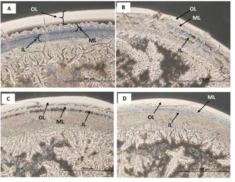
The statistical analysis showed that the outermost layer of the 11-year-old female was significantly thicker compared to the outermost layer of the tear ferning of the 28-year-old female, 28-year-old male, 34-year-old male, and 42-year-old female participants. The outermost layer of the 28-year-old female was thinner than the 42-year-old female, 28-year-old male, and 34-year-old male participants. There was no significant difference between the middle and inner layers among the subjects (Tables 1 and 2).
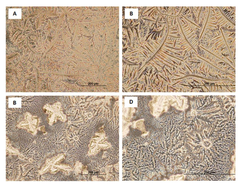
3.1. Ferning Pattern in the Center and Periphery of the Dry Tear Film Droplet
In the 11-year-old female, the branching was very fine and there was no spacing between the branches. The primary branches shot out secondary and tertiary branches which were very thin and distinct from each other. The branching was categorized as Grade 0 (Masmali), Type 1 (Rolando) (Fig. 2A, B). The branching was not very well distinguished, and a star-shaped structure was present in the periphery as well as at the center. The ferning was categorized as Grade 3 (Masmali), Type 4 (Rolando) (Fig. 2C, D). The primary and secondary branching of the 28-year-old male was distinctive, but it had minor spacing between the branching. It was considered as Grade 1 (Masmali), Type 2 (Rolando). In the 34-year-old male and 40-year-old female, the branching had a wider spacing between the secondary and tertiary branches and was graded as Grade 3 (Masmali), Type 4 (Rolando) (Fig. 2C-2F). The ferning pattern in the center and periphery were similar to each other in all subjects.
| Participants | Thickness (µm) | |||
|---|---|---|---|---|
| 1st Layer | 2nd Layer | 3rd Layer | Grand Average | |
| 11-year-old female | 1054.84±10.67 *** | 601.65±9.55 | 676.83±10.58 | 777.77±17.55 |
| 28-year-old female | 538.90±11.54 ***, **, ††† | 572.44±15.85 | 557.51±11.12 | 556.28±7.55 |
| 28-year-old male | 765.82±17.32 ***, ††† | 548.59±15.38 | 792.16±17.92 | 702.19±13.31 |
| 32-year-old male | 730.13±11.38 *** | 632.65±16.65 | 879.81±17.06 | 747.53±12.15 |
| 34-year-old male | 519.26±9.64 ***, ††† | 558±12.18 | 738.18±12.05 | 605.34±10.29 |
| 42-year-old female | 565.70±6.99***, ** | 341.67±4.10 | 642.03±8.58 | 516.47±11.35 |
| Within ♀ | ||
|---|---|---|
| 11-year-old ♀ & 28-year-old ♀ | 0.000 | *** |
| 11-year-old ♀ & 42-year-old ♀ | 0.000 | *** |
| 28-year-old ♀ & 42-year-old ♀ | 0.004 | *** |
| Within ♂ | ||
| 28-year-old ♂ & 32-year-old ♂ | 0.012 | |
| 28-year-old ♂ & 34-year-old ♂ | 0.000 | *** |
| 32-year-old ♂ & 34-year-old ♂ | 0.000 | |
| Between ♂ & ♀ | ||
| 28-year-old ♀ & 28-year-old ♂ | 0.000 | *** |
4. DISCUSSION
In previous studies, most of the tear film ferning patterns in human and animals were graded according to Rolando and Masmali’s grading systems. According to the Rolando system [6], Type 1 ferning is thin, normal, and uniform. The primary ferns are big, and the branches are closely associated without spaces. Type 2 ferning contains smaller primary ferns with short secondary branching with minute empty spaces among the branches compared with Type 1. Type 3 ferning has small primary ferns without any branches and contains randomly distributed large spaces among the branches, including clusters of mucus. In Type 4, the ferning phenomenon is absent and contains disorganized clusters of mucus. Ronaldo’s grading system was successfully repeated and remains popular for identifying normal and pathological tear ferning [17, 18]. Masmali et al. [5] suggested a new grading scale which involved 5 grades of tear ferning.
Previously, it was reported that camel tear film ferning was surrounded by five homogenous layers containing oily droplets and particles, whereas human tear ferning was surrounded by three layers [16]. In the current pilot study, the tear ferning was also surrounded by three peripheral layers. These layers were well organized and well differentiated in the 11-year-old female subject, whereas they were not as clear in the older adult subjects. Cysteine and methionine are essential parts of the tear proteins, and these proteins are important for the tear mucin [13]. We believe that these proteins and the mucin form the peripheral layers of the tear film. The amino acids, proteins, and mucin vary with respect to age in our samples. These contents seem to be higher in the 11-year-old subject and less so in the older adult subjects.
The present study revealed that the tear film ferning in the 11-year-old female was healthy and categorized as Rolando grade Type 1 (Masmali Type 0), whereas the ferning of the 28-year-old female was Rolando Type 3. The tear ferning pattern among the female subjects directly related to their age. As the age of the female subjects increased, the ferning pattern degenerated and the ferning pattern grading increased. Similar patterns were observed in the male subjects, with the 28-year-old male having better ferning than the 34- and 40-year-old male participants. In comparing the male and female subjects, the grading of ferning in male participants was better than that in female participants. The ferning of the 28-year-old male subject (Type 2, Rolando) was of a remarkably better grade than the 28-year-old female.
The main component for the formation of tear ferning is the electrolyte (Na, K, Cl, Mn) concentration. The organization of the ferning pattern is largely determined by not only the concentration but also the ratio of monovalent sodium ions and potassium ions to divalent calcium and magnesium ions [13, 19-21]. The combination of the protein, mucus, and glycoprotein also plays an important role in the formation of tear ferning, especially mucous, which regulates the surface tension of the tear film to spread widely and quickly [19, 20]. Pearce and Tomlinson [13] suggested that the inhibition of the mucin and proteins helps the formation of tear ferning progress, which is contradictory to previous findings [19, 20]. The degeneration or degrading of tear ferning is mainly due to the hyper-osmolarity [20].
Song et al. [15] assessed the effect of estradiol and testosterone on tears using tear breakup time (TBUT) and Schirmer’s tests. The TBUT and Schirmer’s tests were decreased in the rats who had undergone ovariectomy (OVX). The OVX rats were treated with the systemic administration of estradiol and systemic testosterone administration separately to increase their estradiol and testosterone respectively. The systemic administration of the estradiol further decreased the TBUT, whereas systemic testosterone administration increased TBUT and Schirmer’s test results (producing enough tears). Later, Shen and Ma [22] carried out an assessment of 17β-estradiol on the matrix metalloproteinase (MMPs) in tears of postmenopausal women with dry eye conditions. They showed that high levels of 17β-estradiol increased the matrix MMPs activity in tears of postmenopausal women, presumably causing dry eyes. The dry eye subjects had Grade 3 tear ferning [23], and this dry eye ferning is associated with high levels of 17β-estradiol [22].
Masmali et al. [16] reported that camel tear ferning was better than human tear ferning due to the presence of higher amounts of Cl and K. They believe that Cl and K play an important role in the formation of the tear ferning of camel tears. These elements were less than the Cl and K of the refresh plus, which is used as a tear supplement. The 11-year-old female had the best tear ferning pattern in our present study. This might be due to higher Cl and K as well as a balanced ratio of electrolytes, hyper-osmolarity, and optimal macromolecule concentration. The degradation of tear ferning in the 28- and 45-year-old females may be due to the increase in the hyper-osmolarity and disturbance in the ratio of the ration of monovalent (Na & K) and divalent (Ca & Mn). This could be caused due to the increase in estradiol in the older women. Overall, the tear ferning in the male subjects was better compared to the female subjects, which may be because tear ferning was not affected by the testosterone levels in male subjects, whereas it was affected by the estradiol in the female subjects.
Limitations of the current pilot study are the small sample size and narrow range of age groups. Thus, a future study is required to assess tear ferning in a larger sample size of male and female participants and a wider range of age groups. However, this pilot study provides preliminary results that suggest a possible effect of age and gender on the tear ferning of human tears.
CONCLUSION
The current pilot study showed that irrespective of gender, ferning patterning and grading degenerated and increased, respectively, as the age of the participants increased. Furthermore, the grading of ferning was better in male participants compared to female participants.
LIST OF ABBREVIATIONS
| OL | = Outer layer |
| ML | = Middle |
| IL | = Inner layer |
| TBUT | = Tear breakup time |
| MMPs | = Matrix metalloproteinase |
ETHICS APPROVAL AND CONSENT TO PARTICIPATE
The current study was approved by the local ethics committee of King Saud University Saudi Arabia.
HUMAN AND ANIMAL RIGHTS
No animals were used for studies that are the basis of this research. All the humans were used in accordance with the ethical standards of the committee responsible for human experimentation (institutional and national), and with the Helsinki Declaration of 1975, as revised in 2013 (http://ethics.iit.edu/ecodes/node/3931).
CONSENT FOR PUBLICATION
The written informed consent form was taken from the patients and volunteers.
STANDARDS OF REPORTING
STROBE guidelines were followed.
AVAILABILITY OF DATA AND MATERIALS
The data that support the findings of this study are available within the article.
FUNDING
None.
CONFLICT OF INTEREST
The author declares no conflict of interest, financial or otherwise.
ACKNOWLEDGEMENTS
Declared none.

