All published articles of this journal are available on ScienceDirect.

Clinical Outcomes of Toric Implantable Collamer Lens (T-ICL) and Toric Implantable Phakic Contact Lens (IPCL) for High Myopia with Astigmatism: A Comparative Study
Abstract
Background:
Our study aimed to compare the clinical, visual outcomes, and efficacy of toric Implantable Collamer Lens (T-ICL) and toric implantable phakic contact lens (IPCL) in patients with high myopia and astigmatism over a follow-up period of 6 months.
Methods:
A prospective interventional randomized comparative study included 60 myopic eyes divided into 2 groups, group A including 30 eyes that were implanted with T-ICL, and group B, including 30 eyes that were implanted with toric IPCL. The refractive results, visual acuity, central corneal endothelial cell count, and intraocular pressure (IOP) were evaluated at baseline and at 1 and 6 months post-surgery. Any complications either during or after surgery were assessed.
Results:
In both study groups, the mean central corneal endothelial cell count was significantly decreased after 1 month and improved to reach near pre-operative values after 6 months postoperatively, indicating good lens biocompatibility. A statistically significant increase in IOP was found in both groups during the early follow-up, and a significant decrease after 6 months postoperatively (p=0.036) was reported in group A. A significant reduction in both spherical and cylindrical refractive errors with good predictability was reported in both groups compared with pre-operative values. Regarding the predictability, In T-ICL group (A), the median spherical and cylindrical errors were significantly improved from (-10 D & -4.5 D) pre-operatively to (-0.3 D & - 0.3 D) at the end of 6 months follow up period. Similarly, in the toric IPCL group (B), the median spherical and cylindrical errors were significantly improved from (-11 D & -4.5 D) pre-operatively to (-0.3 D & - 0.3 D) by the end of follow up period. A statistically significant improvement of UCDVA at 6 months postoperatively was found in both groups, as median preoperative LogMAR UCDVA was 1.1 and 1.3 in groups A and B respectively, which was improved to 0.3 in both groups at the end of follow-up period. There were no reported intra- or postoperative complications such as cataract, keratitis, or lens decentration.
Conclusion:
Toric IPCL is a suitable alternative to T-ICL for the management of high myopia with astigmatism, especially in developing countries, as it is cheaper and easier to implant than T-ICL. However, data over longer follow-up periods are needed to confirm its safety and stability.
1. INTRODUCTION
Myopia is the most prevalent refractive error in children and adults around the world, causing defective vision [1]. Myopia correction is required to avoid vision impairment. During the last 2 decades, various surgical procedures have been emerged and approved for treatment of different degrees of myopia. Refractive lens exchange, corneal ablation surgery, and phakic intraocular lens implantation (pIOL) are among these procedures. Ablative corneal operations like; laser-assisted in situ keratomileuses (LASIK), photorefractive keratectomy (PRK), laser-assisted subepithelial keratectomy and epi-LASIK, are not appropriate for correcting higher degrees of myopia because it has a higher risk of postoperative complications and poor visual outcomes [2]. Refractive lens surgery is not a suitable treatment option for patients aged less than 40 years due to a higher incidence of retinal detachment and loss of accommodation [3]. Alternatively, phakic intraocular lens implantation (pIOL) can treat significantly higher degrees of myopia that are beyond the range of refractive corneal surgery. Good visual outcomes, higher efficacy, reversibility, preservation of accommodation, long-term predictability, and stability are among the advantages of this procedure [4, 5]. But as pIOL implantation is an intraocular surgery, it may result in problems such as uveitis, pigment dispersion syndrome, cataract, pupillary block glaucoma, and endophthalmitis [6].
Nowadays, the Implantable Collamer Lens (ICL) (Staar Surgical, Nidau, Switzerland) and the Implantable Phakic Contact Lens (IPCL) (Care Group Sight solutions, India) are the available posterior chamber pIOLs on the market. Due to long-term complications, other PC pIOLs such as the phakic refractive lens (PRL, Zeiss Meditec, Jena, Germany) were phased out of the market [7].
Long-term follow-up has shown that the Visian ICL is a safe and effective treatment choice for moderate to high myopia with astigmatism. However, the financial cost of the implant, particularly in developing nations, is a big issue. The toric IPCL is a novel PC pIOL that may be used for the correction of refractive errors as a cost-effective alternative to the ICL implant, which costs 2.5 times as much as the IPCL implant [8, 9].
Our study aimed to compare the clinical, visual outcomes, and efficacy of T-ICL and toric IPCL in patients with high myopia with astigmatism over a follow-up period of 6 months.
2. MATERIALS AND METHODS
2.1. Study Design
A prospective interventional randomized comparative study was conducted during the period from Sept 2020 to May 2021 at Minia University Hospital and Roaa Eye center. A total of 60 myopic eyes with astigmatism were included and divided into 2 groups, group A included 30 eyes that were implanted with T-ICL, and group B included 30 eyes that were implanted with toric IPCL. An approval from the ethical committee of the Faculty of Medicine, Minia University was obtained (Approval No: 677-9/2020). The study adhered to the tenets of the Declaration of Helsinki, and an informed consent was given by all participants.
The inclusion criteria were: age ≥ 21 years, stable refraction (mean spherical equivalent change is ≤ -0.25 D over 1 year), myopia of more than -8 diopters (D) and less than -18 D with astigmatism up to -6 D, endothelial cell count ≥ 2500 cells/mm2, anterior chamber depth (ACD) ≥ 3 mm and IOP < 21 mmHg, with normal ocular examination. Eyes with previous ocular trauma or surgery, other diseases including keratoconus, cataract, glaucoma, uveitis, patients with autoimmune diseases or diabetic patients were excluded from the study.
The following ophthalmic examinations were performed on all patients:
Evaluation of the visual acuity, both uncorrected distance visual acuity (UCDVA) and best-corrected distance visual acuity (BCDVA), using Snellen charts, values were converted to Logarithm of the minimal angle of resolution (logMAR), fundus examination using Volk 90 D lens (Volk, Mentor, Ohio, USA), cycloplegic refraction using Nidek autoref/keratometer (LS 900, HAAGSTREIT DIAGNOSTICS, Switzerland), measurement of anterior chamber depth (internal ACD, measured from the apex of the posterior corneal surface to the apex of anterior lens capsule), keratometric measurements and pachymetry using Pentacam (Oculus Optikgeräte GmbH, Wetzlar, Germany), Pentacam was used also to exclude cases of keratoconus and ectasia, intraocular pressure (IOP) measurement by Tonopen (Reichert, Inc. USA), central corneal endothelial cell count was assessed by specular microscopy (Tomey EM-3000, Tomey Co, and Japan) and white-to-white (WTW) using a Castroviejo caliper (Ambler Surgical, Germany), which was measured after proper caliper calibration, under topical anesthesia while the patient was seated on the examination chair. Measurement was taken by positioning each tip just beyond the clear corneal borders on the limbus, using the slit lamp biomicroscope.
Refraction, ACD, keratometric measurements, pachymetry, and WTW were the required data for the power calculation with target refraction of emmetropia by modified vertex formula as recommended by the manufacturer. The size of the lens depends on ACD and horizontal WTW.
2.2. T-ICL Design
The ICL is a biocompatible hydrophilic collagen hydroxyethyl methacrylate copolymer with an ultraviolet light filtering chromophore. It's a foldable lens that can be injected through a corneal incision of 3.2 mm or smaller. In 2018, the FDA approved the toric version for correcting myopia with astigmatism. It has the potential to correct myopia and astigmatism up to -18.0 D and -6.0 D, respectively. The V4c, commonly known as the EVO variant, features a 360 um central port (KS-Aquaport) that precludes the necessity for iridectomy or iridotomy with Nd: YAG [10] (Fig. 1).
2.3. Toric IPCL Design
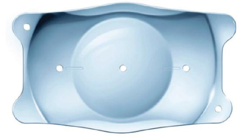
The IPCL is a foldable injectable to be injected through a 2.8 mm corneal incision. It has a unique design with 2 holes in the peripheral part from the upper zone and 4 holes outside the optical zone to facilitate loading in the cartridge and unfolding inside the eye, as well as 6 haptics for added stability. The toric design has the potential to correct myopia up to -30 D and astigmatism up to -10 D. The V2.0 design features a central opening of approximately 380 um, which is aimed to minimize glare and light scattering while also facilitating aqueous circulation, hence alleviating the need for peripheral iridectomy (Fig. 2) [11].

2.3.1. Surgical Procedure
All surgeries were performed under general anesthesia by the same surgeon (M.S). Preoperatively, the pupil was dilated by tropicamide 1% every 10 minutes for a minimum of 30 minutes. To prevent cyclotorsion in the supine position with lens misalignment, the eye was marked with a corneal marker at the horizontal axis at the slit-lamp in the upright position before the surgery. Loading of T-ICL and toric IPCL was done before the corneal incision construction.
2.4. T-ICL
V4c design was used with no need for peripheral iridectomy. A 3.2 mm clear temporal tunnel corneal incision was constructed, with a tunnel length of 1.5 to 1.75 mm parallel to the iris plane. Two side port incisions were made at the 6 and 12 o'clock meridians. Through paracentesis, viscoelastic material (hydroxy propyl methyl cellulose) was injected to partially fill the AC. ICL was injected and allowed to slowly unfold after injection of viscoelastic material. Haptics were tucked under the iris, aligning the toric ICL to the desired axis. To show the degree and direction of rotation from the 0-180 axes, a diagram was given with each lens. Intracameral miotic (Miochol-E: acetylcholine chloride, Bausch & Lomb, Bridgewater, NJ, USA) was injected, then the viscoelastic material was completely irrigated and aspirated, and to ensure that the wounds are watertight, proper hydration of the main wound and side port incisions was performed.
2.5. Toric IPCL
V2.0 design was used with no need for peripheral iridectomy. The cartridge was opened and filled with saline and viscoelastic material. The IPCL was gently held from the container near the haptic using McPherson forceps. The IPCL was placed in the cartridge after detection of its orientation and the cartridge was put in the injector.
A 2.8 mm clear temporal tunnel corneal incision, parallel to the iris plane was constructed, ranging from 1.5 to 1.75 mm for tunnel length. Two side port incisions were made at the 6 and 12 o'clock meridians, and the anterior chamber was partially filled with viscoelastic material (hydroxy propyl methyl cellulose) injected through the paracentesis. The IPCL was slowly injected into the AC for detection of the orientation. The four footplates were placed under the iris plane. Detection of the proper orientation of the toric IPCL in the horizontal axis using the landmarks on the surface of the IPCL was designed to be placed in 0-180 axis. Intracameral miotic (Miochol-E: acetylcholine chloride, Bausch & Lomb, Bridgewater, NJ, USA) was injected, then careful removal of the viscoelastic material through proper irrigation and aspiration was done. Water-tight wounds were then ensured by hydrating the main wound and side ports.
Postoperatively, topical antibiotic as Moxifloxacin HCL 0.5% and topical steroid as Prednisolone acetate 0.5% were used and gradually tapered over 1 month. Topical antiglaucoma eye drops were used as needed in some cases.
UCDVA, BCDVA, cycloplegic refraction, IOP, central endothelium cell count, slit-lamp examination for anterior segment evaluation and adverse effects were recorded at 1 and 6 months postoperatively
2.6. Statistical Analysis
The collected data were coded, tabulated, and statistically analyzed using SPSS program (Statistical Package for Social Sciences) software version 25.
Descriptive statistics were done for parametric (normally distributed) quantitative data by mean, Standard deviation (SD) and minimum and maximum range and for non-parametric quantitative data by median and interquartile range (IQR), while for qualitative data by frequency and percentage.
Distribution of the data was done by Shapiro Wilk test. Analyses were done between the two groups for parametric quantitative data using Independent Samples T-test and for non-parametric quantitative data using Mann Whitney test.
Analyses were done between the two times within the same group for parametric quantitative data using Paired Samples T test and for non-parametric quantitative data using Wilcoxon’s signed rank test.
Analyses were done between the two groups for Qualitative data using Chi Square test.
The level of significance was taken at p-value ≤ 0.05.
3. RESULTS
Thirty eyes in group A were implanted with T-ICL, while 30 eyes in group B were implanted with toric IPCL. Patients demographic data are summarized in Table 1.
Both spherical and cylindrical errors were significantly reduced during the follow up period in both groups, without statistically significant difference between the two groups either pre-operatively or during the follow-up period (Table 2).
A statistically significant improvement of LogMAR UCDVA was observed in both groups during the follow-up period. While, LogMAR BCDVA showed non-statistically significant change during the follow-up period (Table 3).
A statistically significant increase in IOP was found in both groups during the early follow up, and a significant decrease after 6 months postoperatively (p=0.036) was reported in group A (Table 4).
| Group A (T-ICL) | Group B (T IPCL) | P-value | ||
|---|---|---|---|---|
| N=30 | N=30 | |||
| Age (year) | Range Mean ± SD |
(21-38) 27.4±6.1 |
(21-36) 27.1±5 |
0.819 |
| Sex | Male Female |
16 (53.3%) 14 (46.7%) |
20 (66.7%) 10 (33.3%) |
0.292 |
| Spherical error | Group A (T-ICL) | Group B (T IPCL) | P-value | |
|---|---|---|---|---|
| N=30 | N=30 | |||
| Preoperative | Median IQR |
- 10 (-8: - 12) |
- 11 (-9.5: - 14) |
0.101 |
| 1 month postoperatively | Median IQR |
- 0.5 (-0.3: -0.8) |
- 0.5 (-0.5: - 0.8) |
0.272 |
| 6 months postoperatively | Median IQR |
- 0.3 (-0.3: - 0.5) |
- 0.3 (-0.3: - 0.5) |
0.738 |
| P-value versus preoperative | ||||
| 1 m vs pre | <0.001* | <0.001* | ||
| 6 m vs pre | <0.001* | <0.001* | ||
| Cylindrical error | ||||
| Preoperative | Median IQR |
- 4.5 (-4: - 5) |
- 4.5 (-4: - 5) |
0.808 |
| 1month postoperatively | Median IQR |
- 0.8 (-0.3: - 0.8) |
- 0.5 (-0.5: - 0.8) |
0.516 |
| 6 month postoperatively | Median IQR |
- 0.3 (-0.3: - 0.5) |
- 0.3 (-0.3: - 0.5) |
1 |
| P-value versus preoperative | ||||
| 1 m vs pre | <0.001* | <0.001* | ||
| 6 m vs pre | <0.001* | <0.001* | ||
| LogMAR BCDVA | Group A (T-ICL) | Group B (T IPCL) | p-value | |
|---|---|---|---|---|
| N=30 | N=30 | |||
| Preoperative | Median IQR |
1.1 (1: 1.3) |
1.3 (1: 1.3) |
0.270 |
| 1 month postoperative | Median IQR |
0.3 (0.2: 0.4) |
0.3 (0.3: 0.4) |
0.950 |
| 6 months postoperative | Median IQR |
0.3 (0.2: 0.3) |
0.3 (0.2: 0.3) |
0.495 |
| P-value versus preoperative | ||||
| 1 m vs pre | <0.001* | <0.001* | ||
| 6 m vs pre | <0.001* | <0.001* | ||
| LogMAR BCDVA | ||||
| Preoperative | Median IQR |
0.2 (0.2: 0.3) |
0.2 (0.2: 0.2) |
0.379 |
| 1 month postoperative | Median IQR |
0.2 (0.2: 0.3) |
0.2 (0.2: 0.3) |
0.403 |
| 6 months postoperative | Median IQR |
0.2 (0.2: 0.2) |
0.2 (0: 0.2) |
0.418 |
| P-value versus preoperative | ||||
| 1 m vs pre | 0.957 | 0.058 | ||
| 6 m vs pre | 0.263 | 0.197 | ||
| IOP | Group A (T-ICL) | Group B (T IPCL) | P-value | |
|---|---|---|---|---|
| N=30 | N=30 | |||
| Preoperatively | Range Mean ± SD |
(11-18) 14±1.8 |
(12-15) 13.8±1.1 |
0.559 |
| 1 month postoperative | Range Mean ± SD |
(12-19) 15.1±2.1 |
(11-19) 15.3±2.3 |
0.727 |
| 6 months postoperative | Range Mean ± SD |
(11-15) 13±1.3 |
(10-19) 14.1±2.7 |
0.059 |
| P-value versus preoperative | ||||
| 1 m vs pre | 0.017* | 0.001* | ||
| 6 m vs pre | 0.036* | 0.564 | ||
| Central endothelial cell count | Group A (T-ICL) | Group B (T IPCL) | P-value | |
|---|---|---|---|---|
| N=30 | N=30 | |||
| Preoperatively | Range Mean ± SD |
(2980-3322) 3140.6±118 |
(3070-3290) 3154.5±77.4 |
0.591 |
| 1 month postoperative | Range Mean ± SD |
(2900-3200) 3057.7±100.6 |
(2800-3210) 3020.7±113.8 |
0.187 |
| 6 months postoperative | Range Mean ± SD |
(2975-3275) 3135.1±100 |
(2925-3290) 3130.8±95.3 |
0.866 |
| P value versus preoperative | ||||
| 1 m vs pre | <0.001* | <0.001* | ||
| 6 ms vs pre | 0.564 | 0.137 | ||
In both study groups, the mean central corneal endothelial cell count was significantly decreased after 1 month and improved to reach near pre-operative values after 6 months postoperatively (Table 5).
4. DISCUSSION
The prevalence of high myopia with astigmatism is high in Egypt. In a study conducted on 3442 students in their first academic year at Assiut University, Upper Egypt, refractive error was found in 360 of them. Compound myopic astigmatism was found in 257 out of 360 (71.39%) [12].
A large number of myopic persons have been able to improve their quality of life by removing their glasses or contact lenses since the introduction of corneal ablation surgery. Regardless of the fact that current photorefractive corneal surgeries have been demonstrated to be predictable and safe, they are still not appropriate for patients with thin corneas or high myopia [13].
In these cases, phakic IOLs are an acceptable treatment choice since they have a greater ability to correct higher degrees of refractive errors than LASIK and have no risk of corneal ectasia. pIOLs could be used in patients with stable keratoconus after corneal cross-linking [14, 15].
T-ICL is an FDA approved pIOL for the correction of myopia with astigmatism and many studies have confirmed the safety of T-ICL [16-21]. But the cost-effective burden of T-ICL is considered a major concern in our locality. The toric IPCL is a good alternative implant for the correction of high myopia with astigmatism.
Our study aimed to compare the efficacy and safety of both implants (T-ICL and Toric IPCL) over 6 months follow-up period. To our knowledge, this is the first study to evaluate the clinical outcomes of toric IPCL for correction of high myopic astigmatism over 6 months follow-up period.
The current study included 60 myopic eyes, half of them were implanted with T-ICL and the other half were implanted with toric IPCL. Included patients were age-matched (age ≥ 21 years), had stable refraction with normal ocular examination, except for high myopia with astigmatism, who were unfit for keratorefractive surgeries. The included patients had myopia less than -18 D and astigmatism less than -6 D, as T-ICL could correct up to -18 D myopia and -6 D astigmatism while toric IPCL could correct up to -30 D myopia and -10 D astigmatism.
The primary efficacy outcome for the current study is the significant reduction in both spherical and cylindrical refractive errors with good predictability in both groups compared with preoperative values. In T-ICL group (A), the median spherical and cylindrical errors were significantly improved from (-10 D & -4.5 D) pre-operatively to (-0.3 D & - 0.3 D) at the end of a 6-month follow up period (Table 2). Similarly, in the toric IPCL group (B), the median spherical and cylindrical errors were significantly improved from (-11 D & -4.5 D) pre-operatively to (-0.3 D & - 0.3 D) by the end of follow up period (Table 2). Good predictability was achieved with both types of implants, as the mean residual spherical and cylindrical errors were (-0.3 D & - 0.3 respectively) at the end of follow-up period. Similar results were obtained from Sachdev et al. in their comparative study on two different implants (ICL, V4c and IPCL, V1), as residual spherical equivalent error ranged between 0.5 D and 1.0 D of the attempted correction in both groups, at the 12 months postoperative visit, although their study included toric and non-toric implantation with different implants designs [22].
A statistically significant improvement of UCDVA at 6 months postoperatively was another efficacy parameter for the two implants, as median preoperative LogMAR UCDVA was 1.1 and 1.3 in the ICL and IPCL groups, which was significantly improved to 0.3 in both groups at the end of follow-up period (Table 3). Similar results were reported from a study on two toric implants (Eyecryl Phakic Toric IOL or Visian Toric ICL) for myopic astigmatism correction. There was no significant difference between both groups along the 2 years follow up [23].
Both used pIOLs, in the current study, V4c T-ICL and IPCL V2.0 toric were implanted with no need for preoperative iridotomy or intraoperative peripheral iridectomy, as IPCL V2.0 TORIC was designed with a unique central hole (380 μm) in the optic for proper aqueous flow. V4c T-ICL, also has a central hole (360 μm) with two additional holes outside the optic helping aqueous outflow. In a previous study by Kawamorita et al., who evaluated aqueous humor dynamics in patients implanted with V4c model, they reported that the presence of a central hole in Hole-ICLs was beneficial in preventing cataract development due to sufficient aqueous diffusion and nutrients reaching the anterior surface of the lens that maintains adequate lens nutrition [24]. This could be applied to current study results, as no eye in both groups developed cataract by the end of the follow-up period. Although cataract formation was the major complication of pIOLs in many previous studies, that could be referred to as longer follow-up periods and different pIOL designs.[10 & 25]
Over the 6 months follow-up period, IOP was maintained below 21 mm Hg in both groups, and no significant difference was reported between the two groups concerning IOP stability, which could be ascribed to the presence of a central hole within the optic of the two implants that maintained proper aqueous flow, reducing the possibility of the pupillary block and pigmentary glaucoma. A mild statistically significant increase in the IOP was reported in the first month postoperatively in both groups (p-value = 0.017 and 0.001, respectively), possibly due to some retained viscoelastic material, postoperative inflammation, or steroid-induced glaucoma which is more common in highly myopic patients, and was managed by topical antiglaucoma medications, this was in agreement with previous study reports . [26]
Though the materials of both toric pIOLs used in this study were different, as T-ICL is manufactured from Collamer®, a proprietary hydroxyethyl methacrylate (HEMA)/porcine-collagen-based biocompatible polymer material, while T-IPCL is formed from a reinforced hybrid hydrophilic acrylic material, both lenses showed good biocompatibility with intraocular tissues. Lens biocompatibility was assessed in our study by two factors: effect on corneal endothelium and the incidence of adverse events (intense postoperative inflammation, hypopyon, or hyphema). Regarding the effect on the corneal endothelium, we observed a similar effect on the corneal endothelium for both lenses, as the central endothelial cell count was significantly reduced in the early postoperative period, possibly due to surgical trauma, and returned to near preoperative values after 6 months (Table 5). This agreed with results from previous studies held on ICL [27] and IPCL [28]. Sachdev et al. also reported no effect on ECD for both lenses; T-ICL (V4c) and toric IPCL (V1) over a 1-year follow-up period [22].
In current study, none of the eyes demonstrated excessive postoperative inflammatory reaction (endophthalmitis or hypopyon) or hyphema in both groups, and topical steroids were tapered gradually during the first month postoperatively, which indicates good compatibility for both implants. No cases of lens rotation or cataract were recorded during the follow up period.
Toric IPCL has some advantages over T-ICL in terms of surgical technique. We found that toric IPCL loading was much easier than T-ICL loading, which requires special instrumentation and a learning curve. For the implantation of both lenses, a temporal corneal incision was constructed, but the T-ICL had a longer incision length (3.2 mm) than the toric IPCL (2.8 mm), which could impact the final cylindrical error results due to induced astigmatism. The T-ICL had a diagram for axis detection after implantation, while the toric IPCL is customised for a 0-180 degree axis with no rotation. Toric IPCL can also correct a broader range of compound myopic astigmatism (myopia up to-30 D and astigmatism up to-10 D) than T-ICL (myopia up to-18 D and astigmatism up to-6 D).
Limitations to the current study are: the smaller sample size, the shorter follow up period, as longer follow up period may highlight certain adverse effects (ex; decentration), and finally lack of vault evaluation by ultrasound biomiscroscopy (UBM).
CONCLUSION
In conclusion, Toric IPCL provided refractive correction, stability, and visual acuity improvement comparable with T-ICL results for correction of high myopia with astigmatism over 6 months follow-up period, with good biocompatibility. Toric IPCL is a suitable alternative to T-ICL for the management of high myopia with astigmatism, especially in developing countries. However, data over longer follow-up periods are needed to confirm its safety and stability.
LIST OF ABBREVIATIONS
| T-ICL | = Toric Implantable Collamer Lens |
| IPCL | = Toric Implantable Phakic Contact Lens |
| IOP | = Intraocular Pressure |
| LogMAR | = Logarithm of the Minimal Angle of Resolution |
| PRK | = Photorefractive Keratectomy |
| LASIK | = Laser-Assisted in Situ Keratomileuses |
| pIOL | = Phakic Intraocular Lens |
| ACD | = Anterior Chamber Depth |
| BCDVA | = Best-Corrected Distance Visual Acuity |
| UCDVA | = Uncorrected Distance Visual Acuity |
| WTW | = White-to-White |
| SPSS | = Statistical Package for Social Sciences |
| SD | = Standard Deviation |
| IQR | = Interquartile Range |
| HEMA | = Hydroxyethyl Methacrylate |
| UBM | = Ultrasound Biomiscroscopy |
ETHICS APPROVAL AND CONSENT TO PARTICIPATE
The study had gained approval from Local Research Ethics Committee, Faculty of Medicine, Minia University Hospitals, Egypt, with approval No: 677-9/2020.
HUMAN AND ANIMAL RIGHTS
No animals were used that are the basis of this study. All the human experiments were followed in accordance with the tenets of the Helsinki Declaration. Study registration number is: UMIN000043395 (retrospectively registered).
CONSENT FOR PUBLICATION
Written consent form was signed by all participants in the study.
STANDARDS OF REPORTING
STROBE guidelines were followed.
AVAILABILITY OF DATA AND MATERIALS
Data are available from the corresponding author [H.R.A.] on reasonable request.
FUNDING
None.
CONFLICT OF INTEREST
The authors declare no conflict of interest financial or otherwise.
ACKNOWLEDGEMENTS
Declared none.

