All published articles of this journal are available on ScienceDirect.

Real-World Outcomes of Supraciliary MINIject Implantation in Glaucoma: A Single-Centre Retrospective Study

Abstract
Introduction
The MINIject is a device designed for implantation into the supraciliary space to lower intraocular pressure. We report the outcomes up to two years of MINIject implantation in a cohort of Irish patients.
Materials and Methods
A retrospective, single-centre cohort study was conducted through review of all medical records relating to patients who received a MINIject implant between November 2023 and November 2025.
Results
There was a significant reduction in IOP at week 1 and month 1, of 4.4 mmHg and 6.3 mmHg respectively, although the prespecified six-month IOP outcome did not reach statistical significance. The number of IOP-lowering medications was significantly reduced at all timepoints up to year 2. The success rate, defined as IOP ≤21 and > 5 mmHg with a minimum of 20% reduction from baseline, was 21.4% for complete (unmedicated) success, and 35.7% for overall (medicated or unmedicated) success. 15% of devices were malpositioned, and 10% of eyes subsequently required filtration surgery.
Discussion
In this study, we report the first Irish cohort of patients receiving MINIject implants and, to our knowledge, only the fourth real-world series reporting on MINIject to date. Overall, our results are more modest than the results reported in the landmark studies for this device.
Conclusion
An early reduction in IOP was observed following MINIject implantation. It also decreased the number of IOP-lowering medications required, with a large proportion of eyes remaining drop-free. However, there was also a high number of reoperations required for malpositioned devices, which occurred among the earlier procedures in this series. This may suggest a possible learning-curve effect, although this observation should be considered only hypothesis-generating.
1. INTRODUCTION
Glaucoma is a leading cause of irreversible vision loss worldwide [1]. It is a progressive optic neuropathy which results in visual field defects and, in a significant proportion, permanent visual impairment [2, 3]. Raised Intraocular Pressure (IOP) remains the only modifiable risk factor for delaying or preventing disease progression. While most glaucoma is treated with topical eye drops or laser therapy, a significant proportion of patients are inadequately controlled with these treatments and require surgical management. Traditional filtration surgery to lower IOP is often technically challenging and accompanied by significant risk of complications [4, 5].
In recent years, new surgical techniques and devices have been developed under the broad term “Minimally Invasive Glaucoma Surgery” (MIGS) [6, 7]. These comprise various ab-interno approaches to lower IOP, often combined with cataract surgery. While they do not, in general, lower IOP to the same degree as filtration surgery, they tend to have a favourable safety profile, and often enable patients to reduce reliance on topical IOP-lowering medications [8]. Many of these MIGS procedures target the trabecular outflow pathway. More recently, the supraciliary (anterior suprachoroidal) space has emerged as an attractive alternative [9, 10]. The suprachoroidal space is a potential space between the choroid and the internal aspect of the sclera, and the supraciliary space is the term used for the anterior portion of the suprachoroidal space. This space can provide a route for aqueous drainage which bypasses the trabecular meshwork [11].
The CyPass Micro-Stent was the first approved ab-interno device which utilised this space [12]. It demonstrated clinically meaningful lowering of IOP; however, longer-term follow-up showed an increased rate of corneal endothelial cell loss, leading to its voluntary withdrawal from the market [13]. Subsequent analyses suggested that factors that contributed to this complication included device design, depth of implantation, and proximity to the corneal endothelium, underscoring the importance of implant biocompatibility, flexibility, and stable positioning [14].
MINIject® (iSTAR Medical) is a soft, porous, 5mm implant made from proprietary iSTAR material [15, 16]. It is designed to be deployed ab interno into the supraciliary space. Its design is intended to promote biointegration and reduce tissue reaction and fibrosis. At present, it is the only commercially available implant that targets the supraciliary outflow pathway. It can be implanted during cataract surgery or as a standalone procedure.
The efficacy of this device was demonstrated in the STAR-I, II, and III trials, which were prospective, single-armed, and non-randomised [15, 17]. A recent pooled meta-analysis of these studies showed a reduction in IOP of nearly 40% from baseline at two-year follow-up [18]. However, real-world data are lacking. The first UK series to be published did not report as large a reduction in IOP at their six-month follow-up, but still achieved significant success rates [19].
We report the results of MINIject implantation at a single centre by two surgeons experienced in MIGS.
2. MATERIALS AND METHODS
We conducted a retrospective, single-centre, cohort study by reviewing medical records of all patients who received a MINIject implant at Mater Misericordiae University Hospital, Dublin, Ireland between November 2023 and November 2025. All surgeries were performed by one of two surgeons (ED and PA) with a subspecialty interest in glaucoma and MIGS. We used anonymised, retrospective data for the analysis, and the study did not involve intervention or direct patient contact. The Research Ethics Committee of the Mater Misericordiae University Hospital determined that this project constituted a service evaluation rather than research and therefore fell outside the committee’s remit. Formal ethical approval was not required. The analytical dataset was anonymised during extraction: medical record numbers were not included, and no linkage key was retained. No formal Data Protection Impact Assessment (DPIA) was conducted. The study was conducted in accordance with the principles of the Declaration of Helsinki. This study is reported in accordance with the STROBE guidelines for observational studies [20].
2.1. Inclusion Criteria
Consecutive patients who received the MINIject implant were identified through theatre records, patient electronic medical records, and hospital administrative records. Eligibility required receipt of a MINIject implant during the study period. No a priori exclusion criteria were specified, and no eligible patients or eyes were excluded. All patients who had MINIject implantation were included, regardless of glaucoma type or baseline characteristics. The inclusion of all glaucoma subtypes reflects real-world use of the device, while recognising that this may introduce heterogeneity in treatment response. We included cases that were combined with cataract surgery as well as standalone implantation. The study size was the totality of patients who underwent this procedure. No formal sample-size calculation was performed because the study included all consecutive eligible cases during the study period. The decision to perform MINIject implantation was based on the judgement of the operating surgeon without pre-defined criteria. In general, patients with mild-to-moderate glaucoma were selected who required additional IOP lowering or medication reduction, as is common for MIGS procedures.
2.2. Data Collection
Pre-operative (baseline) data were taken from the final visit prior to surgery. IOP was measured at each visit with Goldmann applanation tonometry by an ophthalmologist. Visual acuities, which were recorded in Snellen notation, were converted to logMAR for analysis. We did not routinely measure endothelial cell count; however, previous studies have not demonstrated a significant reduction beyond that expected with cataract surgery alone [18]. There was no fixed schedule for follow-up, and the interval between visits was left to the surgeon’s discretion. Patients were asked to stop their IOP-lowering drops in the immediate post-operative period to get an adequate gauge of unmedicated IOP. Medications were re-introduced at the surgeon’s discretion, taking into account the patient’s individual target IOP and severity of glaucoma. This deliberate cessation of medication may have influenced medication-count outcomes at early postoperative visits.
2.3. Surgical Implantation Technique
All surgeries were conducted under subTenon’s anaesthesia with 3-5mL of a 50:50 solution of lidocaine 2% and bupivacaine 0.5%, with or without hyaluronidase 150 IU/mL as per surgeon’s preference. Surgery was performed from a temporal approach through a 2.2mm clear corneal incision. If combined with cataract surgery, the cataract extraction and Intraocular Lens (IOL) implantation was performed in the usual manner. After insertion of the IOL, the patient’s head was tilted 30 degrees away from the surgeon and the operating microscope was also tilted 30 degrees towards the patient. Visualisation of the angle was achieved with a direct surgical gonio-prism. Cohesive viscoelastic (Healon GV Pro 1.8% sodium hyaluronate) was used to create a cyclodialysis cleft in the nasal or superonasal quadrant of the angle. A cyclodialysis cleft is a disinsertion of the ciliary body from the scleral spur, more commonly seen in trauma, but here created iatrogenically for introducing the device. The device was delivered through this cleft under direct visualisation and advanced until the green collar mark on the implant was at the level of the scleral spur.
2.4. Outcome Measures
The primary outcomes were intraocular pressure and use of IOP-lowering medications at 6 months. Secondary outcomes were IOP and IOP-lowering medications at week 1, month 1, month 3, month 12, and month 24, and surgical success. Surgical success was defined as IOP ≤21 and >5 mmHg with a minimum of 20% reduction from baseline, as previously used in the STAR-I, II, and III studies [17]. Complete success was defined as meeting these criteria without IOP-lowering medication, qualified success as meeting these criteria with IOP-lowering medication, and overall success as either complete or qualified success.
2.5. Statistical Analyses
Descriptive statistics were used to summarise baseline demographic and clinical characteristics. Changes in IOP and number of IOP-lowering medications over time were analysed using linear mixed-effects models, with time included as a categorical fixed effect and patient as a random intercept to account for within-patient correlation among observations. Initial models additionally included a random intercept for eye nested within patient. However, the eye-level variance was estimated at approximately zero for both outcomes, resulting in singular fits, and this term was therefore omitted from the final models.
The models were fitted using restricted maximum likelihood, with degrees of freedom estimated using the Satterthwaite approximation. All available observations were included without imputation of missing values. Estimated marginal means and 95% confidence intervals were calculated at each time point, and postoperative measurements were compared with baseline using Dunnett-adjusted contrasts. The exploratory POAG–NTG comparison additionally included glaucoma subtype and its interaction with time as fixed effects. For this analysis, time-specific between-group comparisons were adjusted using the Holm method. A p-value <0.05 was considered statistically significant. All statistical analyses were performed using R version 4.6.1 (R Foundation for Statistical Computing, Vienna, Austria), with the lme4 (version 2.0-6), lmerTest (version 3.2-1), and emmeans (version 2.0-4) packages. Model diagnostics were performed using the performance (version 0.17.1) and DHARMa (version 0.5.0) packages.
3. RESULTS
20 eyes of 13 patients were identified who had a MINIject procedure. The mean age was 72 ± 8.8 years. 53.8% (7/13) of patients were female, and 46.2% (6/13) were male. Four patients (30.8%) were of Black African ethnicity; the remainder (69.2%) were white. The number of right eyes was 12/20 (60%), and the number of left eyes was 8/20 (40%).
The diagnosis was Primary Open-Angle Glaucoma (POAG) in 14 eyes (70%), Normal-Tension Glaucoma (NTG) in 4 eyes (20%), and Angle-Closure Glaucoma (ACG) in 2 eyes (10%). Only two eyes (10%) had standalone MINIject performed (both eyes already pseudophakic); in the remainder of the eyes (90%), the procedure was combined with cataract surgery (Table 1).
| Mean | ± SD | |
|---|---|---|
| Age (years) | 72 | ± 8.8 |
| BCVA (logMAR) | 0.29 | ± 0.17 |
| Sex | n | (%) |
| Male | 6 | 46.1% |
| Female | 7 | 53.8% |
| Eye | n | (%) |
| Right | 12 | 60% |
| Left | 8 | 40% |
| Ethnicity | n | (%) |
| Caucasian | 9 | 69.2% |
| Black African | 4 | 30.8% |
| Diagnosis | n | (%) |
| POAG | 14 | 70% |
| NTG | 4 | 20% |
| PACG | 2 | 10% |
| Surgery performed | n | (%) |
| MINIject and phacoemulsification | 18 | 90% |
| Standalone MINIject | 2 | 10% |
Abbreviations: BCVA best-corrected visual acuity, logMAR logarithm of the minimum angle of resolution, NTG normal tension glaucoma, PACG primary angle closure glaucoma, POAG primary open angle glaucoma, SD standard deviation.
At each attended visit, patients had their Visual Acuity (VA), IOP, and number of medications recorded. The mean baseline IOP was 20.5 ± 7.2 (range 10–40) mmHg (Table 2). The mean number of medications prior to surgery was 2.4 ± 1.3 IOP-lowering agents, including one patient on oral Diamox. Mean pre-operative best-corrected visual acuity was 0.29 ± 0.17 logMAR.
| - | Baseline | Week 1 | Month 1 | Month 3 | Month 6 | Year 1 | Year 2 |
|---|---|---|---|---|---|---|---|
| Eyes assessed (n) | 20 | 14 | 13 | 11 | 11 | 6 | 8 |
| IOP (mmHg) [mean (SD)] | 20.5 (7.2) | 13.6 (8.6)* | 14.4 (6.3)* | 17.6 (10.1) | 14.3 (5.0) | 13.2 (3.9) | 15.1 (3.1) |
| IOP reduction (mmHg) [mean (SD)] | - | 4.4 (8.7) | 6.3 (10.6) | 1.6 (10.0) | 3.2 (3.7) | 4.0 (2.6) | 2.3 (4.9) |
| IOP reduction (%) | - | 23.8% | 23.6% | 6.7% | 18.0% | 22.5% | 7.1% |
| Number of medications [mean (SD)] | 2.4 (1.3) | 0.0 (0.0)*** | 0.5 (0.9)*** | 0.9 (1.3)*** | 0.5 (0.9)*** | 0.5 (0.8)** | 0.9 (1.0)** |
| Medication-free eyes (%) | - | 100% | 76.9% | 63.6% | 72.7% | 66.7% | 50% |
Abbreviations: IOP intraocular pressure, SD standard deviation.
Note: * p < 0.05; ** p < 0.01; *** p < 0.001.
The only intraoperative complication recorded was mild bleeding in one patient.
The mean follow-up time was 295 ± 224 days (range 26 to 634 days). 14/20 (70%) had at least six months of follow-up. The number of eyes contributing data at each timepoint is presented in Table 2.
Figure 1 shows box plots of IOP and IOP-lowering medications at week 1, month 1, month 3, month 6, year 1, and year 2. In the primary mixed-effects analysis, the overall effect of time on IOP did not reach statistical significance (F (6,67.29) = 2.19, p = 0.054), and the prespecified endpoint of IOP reduction at six months also did not reach statistical significance after adjustment for multiple comparisons. However, the IOP reductions observed at week 1 and month 1 (Table 2) were statistically significant (Dunnett-adjusted p = 0.016 and p = 0.049, respectively). Although the IOP was lower at every other time point, the difference did not reach statistical significance. The number of IOP-lowering medications decreased significantly over time (F (6,66.16) = 12.50, p < 0.001), with all postoperative comparisons to baseline remaining strongly statistically significant after Dunnett adjustment (week 1 to month 6, p < 0.0001; year 1, p = 0.0017; year 2, p = 0.0027).
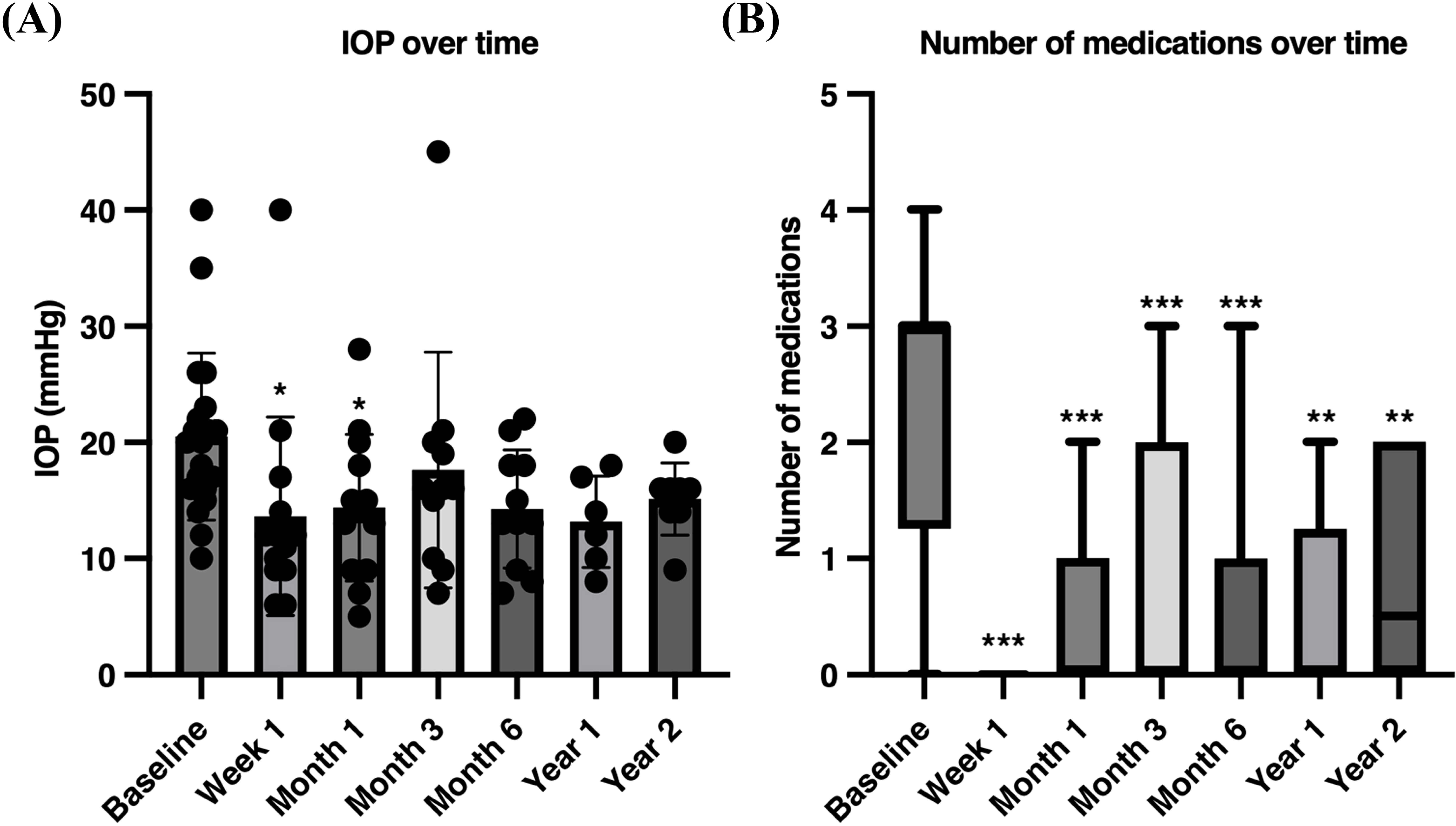
Box plots of intraocular pressure (IOP) (A) and number of medications over time (B).
Note: * p < 0.05; ** p < 0.01; *** p < 0.001.
An exploratory subgroup analysis did not demonstrate a significant difference in the IOP-lowering effect of the procedure between POAG compared with NTG (time x glaucoma subtype interaction, F (6,55.67) = 0.262, p = 0.952). No time-specific POAG-NTG comparison was statistically significant after Holm adjustment (all adjusted p ≥ 0.609; Table S1). However, statistical interpretation was limited by the small number of NTG eyes, and these findings should not be considered evidence of equivalence between glaucoma subtypes.
By the STAR definitions of success (IOP ≤21 and >5 mmHg with a minimum of 20% reduction from baseline), among eyes with at least six months of follow-up, 21.4% (3/14) eyes met the criteria for complete (unmedicated) success. An additional two eyes were qualified (medicated) successes, for a total of 35.7% (5/14) overall success rate.
In terms of post-operative complications, three MINIject devices (15%) were noted to be malpositioned in the post-operative period. In one case, repositioning was attempted at day 7, but due to persistent poor positioning, the implant was removed. A second case underwent successful repositioning at two weeks. In a third patient, the malpositioned MINIject remained under observation only. These malpositioned MINIjects were the second, eighth, and tenth procedures, and there were no poorly positioned implants in the latter half of this cohort. Two eyes (10%) underwent filtration surgery (Preserflo) at a mean of 6 months due to persistent uncontrolled IOP.
4. DISCUSSION
In this study, we report the first Irish cohort of patients receiving MINIject implants and, to our knowledge, only the fourth real-world series reporting on MINIject to date [19, 21-23].
We found a reduction in IOP at all time points compared to baseline, although this was only statistically significant at week 1 and month 1. In the primary mixed-effects analysis, the overall effect of time on IOP did not reach statistical significance. In contrast, the reduction in IOP-lowering medications was statistically significant at every postoperative timepoint. The reduction in medications at the week 1 visit can be attributed to our general policy of asking patients to stop their IOP-lowering medication in the operated eye until review. The proportion of eyes remaining medication-free therefore decreased over time, from 100% at first post-operative review to 50% at year 2 as medications were reintroduced.
Overall, our results are more modest than the results reported in the STAR studies. The mean reduction in IOP in the pooled results from the STAR studies was 9.4 ± 4.7 mmHg at six months, and the rates of overall success (qualified plus complete success) and complete success were 89.4% and 36.4%, respectively, at 24 months [18]. By comparison, our overall success rates were 35.7%, including 21.4% complete success.
In the UK real-world study, success rates were reported as 67.6% overall success and 56.8% complete success [19]. However, they elected to use a different definition of success from the STAR trials, namely IOP ≥ 6 ≤ 18 mmHg, i.e., without a requirement for a percentage reduction. If we had used their definition of success, our overall and complete success rates would be 85.7% and 57.1%, respectively. This highlights the need to scrutinise definitions of success across different studies carefully.
Among possible explanations for our less impressive results is that we included patients with NTG, whereas all patients in the STAR studies required a medicated IOP between 21 and 25 mmHg. While many MIGS procedures have shown efficacy in NTG, the response to MIGS may differ from POAG [24]. In our exploratory subgroup analysis, we found no evidence of a difference in IOP change over time between POAG and NTG. However, only four NTG eyes from two patients were included, resulting in very limited statistical power and imprecise estimates. The absence of significance should therefore not be interpreted as evidence of equivalent treatment effects between the two glaucoma subtypes.
Additionally, the baseline IOP in our cohort was lower, so we might expect to see proportionally less of a reduction in IOP. However, our IOP reduction was also less impressive than the real-world study in the UK, which had a similar baseline IOP and also included normal tension glaucoma [19]. We are unable to account for this difference except that these were non-randomised retrospective studies without defined recruitment criteria or a study protocol, in which patient selection may have varied considerably. Additionally, the UK real-world study was conducted in an entirely Caucasian population, while our population had a significant minority of non-white patients. Glaucoma in these patients is often more severe and may demonstrate different responses to surgical interventions [25].
There was a relatively high level of malpositioned devices (15%), two of which required early reoperation. In another series of patients undergoing MINIject in the context of failed filtration surgery, 2/18 (11.1%) of devices were reported to have luxated into the anterior chamber in the early post-operative period, requiring revision [23]. In the UK series, no malpositioned devices were reported, although there were at least two reoperations, one for hyphaema wash-out, and one to manage aqueous misdirection [21].
Looking at the pooled data from the STAR-I, -II, and -III studies, only one device was noted to have dislocated into the anterior chamber requiring explantation [18]. The STAR-II study reported one case in which the implant was delivered partially in the wrong space, and one device that was implanted too far posteriorly, but no repositioning was reported [17]. In our series, all three malpositioned devices were clustered in the first 10 cases, and there were no malpositioned devices in the latter 10 cases. This may suggest a possible learning-curve effect, but given the small number of procedures and events, this observation should be considered hypothesis-generating only.
There were several limitations to this study. Data were collected retrospectively, and there was no set protocol for visits; therefore, caution is warranted when attempting to extrapolate these results to other populations. Reasons for individual missed visits were not systematically recorded. Although the mixed-effects model allowed inclusion of all available data, this does not eliminate the possibility of attrition bias if eyes with incomplete follow-up differed systematically from the rest of the group. The success analysis was also restricted to eyes with at least six months of follow-up and may therefore be subject to similar attrition bias. Due to this variability in follow-up, estimates at years 1 and 2 should be interpreted with caution, given the smaller number of eyes contributing to the results at these time points. There was no requirement to record any specific complications, so it is possible that intraoperative complications may have been encountered more frequently than recorded (e.g. mild hyphaema) if not deemed clinically significant at the time of surgery.
As there was no control group, we are unable to account for the size of the effect of IOP lowering induced by phacoemulsification alone, or by regression to the mean. Only two eyes underwent standalone MINIject implantation, limiting meaningful comparison between standalone and combined procedures. There was no pre-operative washout of medications, although it is important to note that the STAR studies did not perform washout either. The post-operative management was also not performed according to a particular protocol, and there was no specific target for reduction in IOP. Additionally, we did not routinely collect endothelial cell count, which may have been a useful measurement to record.
CONCLUSION
In this series, eyes that received MINIject implant demonstrated a numerical reduction in IOP across follow-up, with statistically significant reductions at week 1 and month 1 in the primary analysis. There was also a significant decrease in the number of IOP-lowering medications, which was sustained across the two-year follow-up. The proportion of eyes remaining drop-free decreased over time, from 77% at month 1 to 50% by year 2. However, there was also a high number of reoperations required for malpositioned devices, which occurred among the earlier procedures in this series. This may suggest a learning-curve effect, but given the small sample size, this observation should be considered hypothesis-generating only.
AUTHORS’ CONTRIBUTIONS
The authors confirm their contributions to the paper as follows: E.S., P.A., E.D.: Study conception and design; E.S.: Data collection; E.S., P.A., E.D.: Analysis and interpretation of results; E.S.: Draft manuscript. All authors reviewed the results and approved the final version of the manuscript.
LIST OF ABBREVIATIONS
| ACG | = Angle-Closure Glaucoma |
| IOP | = Intraocular Pressure |
| logMAR | = logarithm of the Minimal Angle of Resolution |
| MIGS | = Minimally Invasive Glaucoma Surgery |
| NTG | = Normal Tension Glaucoma |
| POAG | = Primary Open-Angle Glaucoma |
| VA | = Visual Acuity |
ETHICS APPROVAL AND CONSENT TO PARTICIPATE
The Research Ethics Committee of the Mater Misericordiae University Hospital, Ireland determined that this project constituted a service evaluation rather than research and therefore fell outside the committee’s remit. Formal ethical approval was not required.
HUMAN AND ANIMAL RIGHTS
All human research procedures followed were in accordance with the ethical standards of the committee responsible for human experimentation (institutional and national), and with the Helsinki Declaration of 1975, as revised in 2013.
CONSENT FOR PUBLICATION
Informed consent was waived for this retrospective study due to the exclusive use of de-identified patient data, which posed no potential harm or impact on patient care.
AVAILABILITY OF DATA AND MATERIALS
All data generated or analyzed during this study are included in this published article.
ACKNOWLEDGEMENTS
Declared none.