All published articles of this journal are available on ScienceDirect.

Neutral Density Filters as a Tool for Cycloplegic Plusoptix-Photorefractor Measurements: An Explorative Study
Authors Info & Affiliations
Abstract
Purpose:
The purpose of this study is to investigate the usefulness of neutral-density (ND) filters in cycloplegic-Plusoptix-photorefractor measurements.
Methods:
No-filter and ND-filter 0.04, 0.1 and 0.2 cycloplegic-Plusoptix-photorefractor measurements were made in 42 hypermetropic eyes. Sphere, cylinder, spherical equivalent (SEQ), J0, and J45 values were compared.
Results:
Mean Plusoptix-photorefractor pupil sizes were 7.7±0.68 and 7.7±0.72 mm The no-filter failure rate was 16%, with 87% in pupils >7.8 mm. Mean no-filter sphere, cylinder, SEQ, J0 and J45 values were +0.34±0.35D, -0.29±0.22D, +0.20±0.36, -0.00±0.15, and +0.02±0.11, respectively. Only ND-filter-0.04 provided 5% more successful measurements and a clinically significant alteration in the percentage of values exceeding 0.5D for sphere and SEQ (-10% and -20%), but not for cylinder (+5%). Despite the increased accuracy, 21% of the spherical outcome exceeded 0.50D. Furthermore, the single-measure-intraclass-correlation-coefficient between no-filter and ND-filter-0.04 outcome was moderate (sphere 0.78 (0.62-0.87), cylinder 0.59 (0.35-0.75), SEQ 0.68 (0.48-0.82), J0 0.73 (0.54-0.84) and J45 0.57 (0.50-0.86)) and indicated significant individual variation. Bland-Altman-analyses indicated significant bias for sphere and SEQ; p=0.038 and p=0.030.
Conclusion:
ND-filter-0.04 resulted in a larger proportion of successful measurements and an increased accuracy. However, an unacceptable percentage of inaccuracy was still present compared to retinoscopy. There could be validity issues with the ND-filter 0.04 or the baseline no-filter readings at the start. We conclude that cycloplegic Plusoptix-photorefraction, even with the use of a 0.04 ND filter, is not a suitable method for exact objective refraction purposes in children.
1. INTRODUCTION
Retinoscopy in cycloplegia is the most reliable method to assess the refractive state of pediatric patients, but it requires trained examiners. Table mounted autorefractors or handheld devices such as the 2nd and 3rd generation Retinomax are excellent alternatives and can be operated easily. Pediatric studies using Retinomax report a mean difference of less than 0.09 diopters [1, 2]. Although reliable, especially in young children, Retinomax requires good cooperation. Furthermore, due to the proximal position and the monocular setting, outcomes can be strongly influenced by residual accommodation, which can be significant in darker pigmented eyes [3, 4].
The handheld videorefractors of Plusoptix GmbH (Nurnberg, Germany) are very patient-friendly, do not require highly trained staff, and have a smaller effect on accommodation due to their setting at 1 meter. The Plusoptix devices use off-axis eccentric infrared photo-retinoscopy in a binocular setting. At a distance of 1 meter, infrared light of 790 nm is projected into the eye and reflected from the retina. Each refractive error has its specific pattern of reflection and brightness. The Plusoptix software translates this pattern into a refractive value. The limits of measurement include a pupil size between 3.3 and 7.8 mm and a spherical and cylindrical power between +5 and -7 diopters.
The Plusoptix devices are not designed and not recommended for cycloplegic use. There are, however, a few studies that analyzed the performance of Plusoptix devices in cycloplegia. All agreed that spherical values and SEQ were overestimated in cycloplegic Plusoptix measurements [1, 5-8]. For cylindrical values, however, an excellent agreement [6, 7], as well as a poor agreement [1, 5], was found. Schmidt-Bacher et al. [1] obtained 55% successful data in a study with 74 children. The failure rate was 45%, with 24% due to a lack of cooperation, 27% due to refractive limitations, and 49% due to light overexposure in large pupils.
Neutral density (ND) filters, also known as gray filters, decrease the intensity of light without selectively affecting specific wavelengths of light from the source. ND-filters achieve attenuation through reflection; through a metal coating or absorption of light. There are ND filters available to attenuate spectral regions from 250 to 2500 nm. The level of attenuation can be specified from optical density (OD) 0.04 to 4.0. The amount of transmission (T) can be calculated by the following equation: OD=-log(T) or T=10-OD. For OD 0.04, the transmission is 91%, and for OD 4.0, the transmission is 0.01%.
Schmidt-Bacher et al. [1] used an OD 0.5 reflective neutral density filter as a test to diminish the percentage of light reflection in large pupils in their study. This filter, however, decreased the success rate of obtaining data from 72% to 67%. In contrast, the accuracy of the mean SEQ increased from 0.22±0.86D to 0.10±0.70D.
Currently, the Plusoptix devices are used as a non-cycloplegic screening tool. Although cycloplegic measurements are more or less restricted by limitations of the device and the relatively low agreement compared to cycloplegic autorefraction and retinoscopy, keeping in mind its patient friendliness and simplicity of operation, a cycloplegic function with reliable data could be an advantage for the determination of refractive errors. The addition of a specific absorptive ND-filter might perhaps validate the Plusoptix-photorefractors for cycloplegic use. The purpose of this study was to investigate 1) the performance of the Plusoptix-photorefractor in cycloplegia, 2) whether ND-filters change cycloplegic outcomes, 3) whether ND-filters generate more successful cycloplegic measurements and more exact cycloplegic refractive outcomes and 4) the optimal ND-filter.
2. MATERIALS AND METHODS
2.1. Materials
Refractive measurements were made with the Retinomax-K+3 and the PowerRefractor II device using Plusoptix-A09 software. The neutral density filters used were OD 0.04 (Newport Corporation, Irvine, USA), OD 0.1 (Newport Corporation), OD 0.2 (Thorlabs Ltd, Newton, USA), and OD 0.3 (Thorlabs Ltd) transmission absorptive filters. According to the specifications of the manufacturers, the 790 nm wavelength resulted in a 91, 79, 61, and 47% light transmission for ND-filter 0.04, 0.1, 0.2, and 0.3, respectively. However, since the infrared light of the device is attenuated twice, encountering the ND-filter going towards the eye and encountering the ND-filter following reflection from the eye, the real amount of light reaching the device is 83% (ND-filter 0.04), 63% (ND-filter 0.1), 39% (ND-filter 0.2) and 22% (ND-filter 0.3), respectively. The ND filters were fitted in a handheld frame. To determine the general effect of the ND-filters on the refractive outcome, at first, un-dilated, non-cycloplegic measurements were made. Thereafter dilated, cycloplegic measurements were made.
2.2. Subjects
To exclude cooperation issues, we conducted this study on adults. Hypermetropic subjects were chosen since the vast majority of small infants are hypermetropic. To investigate the unbiased, i.e., non-cycloplegic influence, we applied the ND-filters also in a non-cycloplegic state. Non-cycloplegic measurements: Ophthalmic staff aged 35 to 60 years who had full spectacle-corrected hypermetropia and undilated pupil diameters between 3.5 and 7.7 mm. Cycloplegic measurements were made in subjects aged 18 to 60 years with pupil diameters of >6mm that received cycloplegics in accordance with their periodical or annual ophthalmic examination. In both regimes subjects with cataract, vitreous hemorrhage, or abnormally shaped pupils, and subjects with spherical values of >+7.00 diopters (D) or cylindrical values of >-4.00D and SEQ <0.00 were excluded.
2.3. Methods
In both cycloplegic and non-cycloplegic subjects Retinomax-K+3 measurements were made. To be able to investigate the true effect of the neutral density filters and minimize the influence of refractive error, subjects received a trial frame with full Retinomax hypermetropic and astigmatic correction, thereby pursuing emmetropia, during Plusoptix-photorefractor measurements. In non-cycloplegia, subjects adapted to room light for 5 minutes to prevent differences in pupil dynamics during measurements. In cycloplegia, measurements were performed 40 minutes after the first eye drop and immediately after the Retinomax measurements. The subjects up to 50 years received 1 drop of cyclopentolate 1% followed by 1 drop of tropicamide 1% with an interval of 5 minutes. The subjects >50 years received 2 drops of tropicamide 1% with an interval of 5 minutes. The Plusoptix-photorefractor measurements were made according to the set-up of Fig. (1).
Consecutive measurements were made without filter and with a 0.04, 0.1, 0.2, and 0.3 ND-filter. The ND-filters were held in front of the trial frame (Fig. 1). Success (data) or failure (no data) was scored along with pupil size, sphere, cylinder, and axis. Every individual measurement was repeated 3 times and mean values were calculated. In the case of 2 measurement results, the average was calculated. If only 1 result was obtained, this result was used. Plusoptix-A09 software measures pupil sizes up to 1 decimal place. The Plusoptix-photorefraction pupil size values were allocated to either category ≤7.7mm or ≥7.8mm. The pupil size of 7.7mm is a manufacturer described device limitation to conduct photo-refraction. Thereafter data acquisition might be limited and perhaps less accurate. In designing this study it was foreseen that a significant proportion of subjects would have cycloplegic pupils of >7.7mm. Dividing into these 2 categories provides the opportunity to investigate the failure of acquisition in very large pupils and whether the refractive outcome generated differs from the refractive outcome with small pupils.
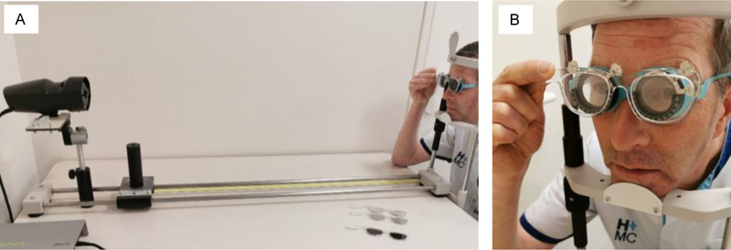
A) subject positioned at a 1-meter distance from Plusoptix-Powerrefractor, wearing a trial frame with full Retinomax-K+3 refractive values. In consecutive order measurements without and with a 0.04, 0.1, 0.2, and 0.3 Neutral Density filter were made.
B) Neutral-Density Filter 0.04 fitted in a handheld frame, positioned in front of the trial frame.
Conversion of a sphero-cylindrical value to an SEQ is a commonly used parameter in pediatric ophthalmology. However, to accurately make statistical comparisons between cylindrical values, it is methodologically necessary to convert the sphero-cylindrical outcomes into Cartesian conversions [9]. According to Fourier decomposition, all sphero-cylindrical outcomes were converted not only in an SEQ value but also into two Jackson cross-cylinder vectors; i.e. J0 and J45 and were admitted in our results and analyses. SEQ, J0 and J45 were calculated with the formulas: sphere+½cylinder (SEQ), -cylinder/2xcos2α (J0) and -cylinder/2xsin2α (J45) respectively [9]. For cycloplegic measurements, mean parameter values were allocated to either category <0.25D, >0.25-0.50D or >0.50D. In designing the study, these categories were chosen to gain more insight into de distributions of the individual refractive outcome; <0.25D can be considered clinically insignificant, and >0.50D as clinically highly relevant. Differences in the distribution in these categories in baseline no-filter and/or the consecutive ND-filters can be of clinical interest.
2.4. Statistics
The number of participants was calculated to detect a mean difference of 0.25D at a minimum between measurements without and with an ND-filter with an alpha of 0.05 and a power of 90%. The standard deviation of difference was set at 0.35D, yielding a sample size of 42 eyes. Outcomes were analyzed with SPSS version 26.
Normality of distribution (Kolmogorov-Smirnov), the intraclass-correlation-coefficient (ICC; absolute agreement, 2-way Mixed-effects model; single measures), and Bland-Altman analyses were made before analyses regarding combining right and left eye outcomes of the sphere, cylinder, J0 and J45 in the analyses. To correct for correlations between right and left eye measurements a Linear Mixed Model approach was used for the difference between baseline Plusoptix-photorefractor outcome and the outcomes of the consecutive ND-filters in both regimes and the influence of pupil size in cycloplegia. The optimal performing ND-filter; regarding the refractive outcome and percentage of successful measurements, as compared to No-filter outcomes. Differences in the distribution of percentages of values within ≤0.25D, >0.25-0.50D, and >0.50D were analyzed using the Wilcoxon Signed Rank Test. Furthermore, the correlation and agreement between outcomes were analyzed using the ICC and Bland-Altman analyses.
Differences were considered statistically significant if p<0.05 and clinically significant if the mean sphere, cylinder, J0, and J45 were >0.25D. Agreement was considered low if ICC was <0.50, moderate if ICC was ≥0.50 to ≤0.75, good if ICC >0.75 to ≤0.90 and excellent if ICC >0.90 [10, 11]. A difference of 5% success rate in obtaining data was considered clinically significant. If the use of a specific filter resulted in less than 70% data acquisition, then this specific filter was not included in the analyses.
2.5. Ethical Considerations
The study was conducted according to the declaration of Helsinki. In the Netherlands, the Medical Research Involving Human Subjects Act (WMO) applies to all medical, scientific research in which humans are subjected to procedures or follow the rules or behavior. Ethical approval by the Institutional Review Board was asked (METC-South West Holland number: 10-077; sub-study number 10-120). Since no burden or risks were involved for the subjects of this study a non-WMO-declaration was received. The study is registered as NL2639/NTR 2767 in the Dutch trial registry.
3. RESULTS
3.1. Non-cycloplegic Measurements
The mean age was 49.2+6.2 years. Retinomax data were obtained in 100% of the measurements. The mean Retinomax SEQ was +1.29±1.43D for the right eye and +1.35±1.48D for the left eye measurements. The mean pupil sizes were 4.4+0.95 and 4.4+0.97mm.
No filter Plusoptix-photorefractor data were obtained in 98% of the measurements. The mean pupil sizes were 5.3±0.77 and 5.5±0.85mm. For ND-filters 0.04, 0.1, 0.2 and 0.3 data were obtained in 98% (ND-filter 0.04), 91% (ND-filter 0.1), 71% (ND-filter 0.2) and 48% (ND-filter 0.3) of the repeated Plusoptix-photorefractor measurements. ND-filter 0.3 was not admitted in analyses or table presentations due to a success rate of <70%. Because of averaging of the repeated measurements 42, 42, 38, and 32 eyes were available for analyses in No filter and ND-filters 0.04, 0.1, and 0.2 respectively. Mean sphere, cylinder, SEQ, J0, and J45, and percentage of values within the categories ≤0.25D, >0.25-0.50D, and >0.50 were admitted in Table 1.
There was a normal distribution for all parameters. The ICC (single measure) between right- and left eye values were: sphere 0.636 (0.292-0.834), cylinder 0.466 (0.055-0.743), SEQ 0.582 (0.212-0.806), J0 0.510 (0.112-0.768) and J45 -0.015 (-0.435-0.411) respectively. Bland-Altman analyses showed no significant agreement between right and left eye measurements in all parameters.
Although the subjects were optically fully corrected, the Plusoptix-photorefractor baseline No-filter measurements showed borderline clinically significant more hypermetropia and clinically significant more mean astigmatism (Table 1). We explored the influence of the individual ND–filters. Compared to the baseline No-filter outcomes, no statistically nor clinically significant changes were present for all parameters in any of the ND-filters (Table 2).
| Mean/SD | No Filter n=42 |
ND-Filter 0.04 n=42 |
ND-Filter 0.1 n=38 | ND-Filter 0.2 n=32 |
|---|---|---|---|---|
| Sphere | +0.25±0.49 | +0.20±0.54 | +0.33±0.68 | +0.42±0.40 |
| Cylinder | -0.40±0.28 | -0.37±0.22 | -0.46±0.31 | -0.43±0.26 |
| SEQ | +0.05±0.51 | +0.02±0.54 | +0.10±0.73 | +0.20±0.42 |
| J0 | -0.04±0.15 | -0.04±0.13 | -0.04±0.18 | -0.07±0.17 |
| J45 | -0.02±0.21 | 0.00±0.17 | -0.02±0.21 | -0.01±0.17 |
| Mean Sphere | Diff | SE | 95% CI | p | Mean Cylinder | Diffa | SE | 95% CI | p | ||||||
|---|---|---|---|---|---|---|---|---|---|---|---|---|---|---|---|
| ND-filter 0.2 | +0.16 | 0.10 | -0.05 / +0.41 | 0.128 | ND-filter 0.2 | -0.02 | 0.06 | -0.13 / +0.10 | 0.795 | ||||||
| ND-filter 0.1 | +0.07 | 0.13 | -0.19 / +0.34 | 0.585 | ND-filter 0.1 | -0.08 | 0.06 | -0.20 / +0.05 | 0.245 | ||||||
| ND-filter 0.04 | -0.05 | 0.11 | -0.27 / +0.18 | 0.669 | ND-filter 0.04 | +0.02 | 0.05 | -0.08 / +0.13 | 0.684 | ||||||
| No filter | +0.25±0.28D | No filter | -0.40±0.28D | ||||||||||||
| Mean SEQ | Diff | SE | 95% CI | p | Mean J0 | Diff | SE | 95% CI | p | ||||||
| ND-filter 0.2 | +0.15 | 0.11 | -0.06 / +0.36 | 0.167 | ND-filter 0.2 | -0.02 | 0.04 | -0.10 / +0.05 | 0.559 | ||||||
| ND-filter 0.1 | +0.03 | 0.14 | -0.25 / +0.31 | 0.819 | ND-filter 0.1 | +0.00 | 0.04 | -0.07 / +0.08 | 0.915 | ||||||
| ND-filter 0.04 | -0.04 | 0.11 | -0.27 / +0.18 | 0.702 | ND-filter 0.04 | +0.01 | 0.03 | -0.06 / +0.07 | 0.868 | ||||||
| No filter | +0.05±0.51D | No filter | -0.04±0.15D | ||||||||||||
| Mean J45 | Diff | SE | 95% CI | p | ||||||
| ND-filter 0.2 | +0.04 | 0.04 | -0.05 / +0.12 | 0.437 | ||||||
| ND-filter 0.1 | -0.01 | 0.05 | -0.10 / +0.09 | 0.877 | ||||||
| ND-filter 0.04 | +0.02 | 0.04 | -0.07 / +0.10 | 0.669 | ||||||
| No filter | -0.02±0.21D | |||||||||
3.2. Cycloplegic Measurements
The mean age of the cycloplegic group was 27.0+5.7 years. Retinomax data were obtained in 100% of the measurements. The mean SEQ was +1.33±0.76D for the right eye measurements and +1.36±0.95D for the left eye measurements. The mean pupil sizes were 7.4±0.83 and 7.5±0.77mm.
No-filter Plusoptix-photorefractor data were obtained in 84% of the repeated Plusoptix-photorefractor measurements. The mean pupil sizes were 7.7±0.68 and 7.7±0.72mm. For ND-filters 0.04, 0.1, 0.2 and 0.3 data were obtained in 89% (ND-filter 0.04), 84% (ND-filter 0.1), 84% (ND-filter 0.2) and 68% (ND-filter 0.3) of the repeated Plusoptix-photorefractor measurements. The ND-filter 0.3 was not admitted in analyses or table presentations due to a success rate of <70%. In our study, 55% of the eyes had pupil sizes of ≥7.8mm. The general failure of obtaining data acquisition described in the previous paragraph can be almost exclusively; ie in 94.1%, attributed to this specific large pupil group. Because of averaging of the repeated measurement 42, 42, 40, and 36 eyes were available for analyses in No-filter and ND-filters 0.04, 0.1, and 0.2, respectively. The ICC (single measure) between right- and left eye values were: sphere 0.851 (0.668-0.937), cylinder 0.234 (-0.204-0.596), SEQ 0.718 (0.422-0.876),0.582 (0.212-0.806), J0 0.317 (-0.139-0.656) and J45 -0,122 (-0.550-0.333) respectively. Bland-Altman analyses showed no significant relation between right and left eye measurements in all parameters.
The mean sphere, cylinder, SEQ, J0, and J45 and the percentage of values within the categories ≤0.25D, >0.25-0.50D, and >0.50D, are admitted in Table 3. Although optically full corrected, clinically, significant more hypermetropia and astigmatism were present in the baseline No-filter PowerRefractor measurements
Correcting for pupil size showed only for parameter “Cylinder” statistically negatively influenced on the amount of astigmatism for ND-filters 0.1 and 0.2 (Linear Mixed Model), (Table 3). The influence of pupil size is visible for astigmatism; “Cylinder”, “J0” and “J45”, where pupils exceeding 7.7mm were found to statistically influence outcomes (Table 4). However, for both the former and the latter, inspecting the 95% CI indicated that no clinical significance (i.e.>0.25D) can be present.
| No-Filter N=42 |
ND-Filter 0.04 N=42 |
ND-Filter 0.1 N=40 |
ND-Filter 0.2 N=36 |
||
|---|---|---|---|---|---|
| Sphere | Mean/SD | +0.34±0.34 | +0.34±0.28 | +0.47±0.34 | +0.49±0.40 |
| ≤0.25D | 45.2% | 50.0% | 35.0% | 33.3% | |
| >0.25-0.50D | 23.8% | 28.6% | 27.5% | 22.2% | |
| >0.50D | 31.0% | 21.4% | 37.5% | 44.4% | |
| Cyl | Mean/SD | -0.29±0.22 | -0.37±0.21 | -0.39±0.22 | -0.42±0.25 |
| ≤0.25D | 57.1% | 50.0% | 40.0% | 41.6% | |
| >0.25-0.50D | 28.6% | 31.0% | 35.0% | 30.6% | |
| >0.50D | 14.3% | 19.0% | 25.0% | 27.8% | |
| SEQ | Mean/SD | +0.20±0.36 | +0.16±0.25 | +0.27±0.31 | +0.28±0.35 |
| ≤0.25D | 52.4% | 64.3% | 47.5% | 44.4% | |
| >0.25-0.50D | 21.4% | 28.6% | 37.5% | 30.6% | |
| >0.50D | 26.2% | 7.1% | 15.0% | 25.0% | |
| Jo | Mean/SD | -0.00±0.15 | +0.02±0.18 | +0.01±0.19 | 0.00±0.20 |
| ≤0.25D | 92.9% | 88.1% | 82.5% | 83.3% | |
| >0.25-0.50D | 7.1% | 11.9% | 17.5% | 16.7% | |
| >0.50D | - | - | - | - | |
| J45 | Mean/SD | +0.02±0.11 | +0.01±0.11 | -0.01±0.14 | +0.03±0.14 |
| ≤0.25D | 100% | 95.2% | 92.5% | 91.7% | |
| >0.25-0.50D | - | 4.8% | 7.5% | 8.3% | |
| >0.50D | - | - | - | - | |
| Mean Sphere | Diff | SE | 95% CI | p | Mean Cylinder | Diff | SE | 95% CI | p | |||||
|---|---|---|---|---|---|---|---|---|---|---|---|---|---|---|
| ND-filter 0.2 | +0.15 | 0.08 | -0.02 / +0.32 | 0.080 | ND-filter 0.2 | -0.12 | 0.05 | -0.23 / -0.01 | 0.032 | |||||
| ND-filter 0.1 | +0.13 | 0.08 | -0.02 / +0.28 | 0.085 | ND-filter 0.1 | -0.10 | 0.05 | -0.20 / -0.001 | 0.049 | |||||
| ND-filter 0.04 | -0.00 | 0.07 | -0.13 / +0.13 | 0.998 | ND-filter 0.04 | -0.07 | 0.05 | -0.17 / +0.02 | 0.118 | |||||
| No filter | +0.34±0.34D | No filter | -0.29±0.22D | |||||||||||
| Pupil ≥7.8mm | -0.00 | 0.05 | -0.10 / +0.10 | 0.952 | Pupil ≥7.8mm | -0.08 | 0.04 | -0.15 / -0.01 | 0.025 | |||||
| Pupil ≤7.7mm | +0.36±0.30D | Pupil ≤7.7mm | -0.26±0.22D | |||||||||||
| Mean SEQ | Diff | SE | 95% CI | p | Mean J0 | Diff | SE | 95% CI | p | |||||
| ND-filter 0.2 | +0.09 | 0.08 | -0.06 / +0.25 | 0.243 | ND-filter 0.2 | +0.01 | 0.04 | -0.07 / +0.09 | 0.782 | |||||
| ND-filter 0.1 | +0.09 | 0.07 | -0.05 / +0.23 | 0.221 | ND-filter 0.1 | +0.01 | 0.03 | -0.06 / +0.08 | 0.851 | |||||
| ND-filter 0.04 | -0.03 | 0.07 | -0.16 / +0.10 | 0.646 | ND-filter 0.04 | +0.02 | 0.03 | -0.05 / +0.09 | 0.544 | |||||
| No filter | +0.20±0.36D | No filter | -0.00±0.15D | |||||||||||
| Pupil ≥7.7mm | -0.03 | 0.05 | -0.13 / +0.06 | 0.469 | Pupil ≥7.8mm | +0.08 | 0.03 | +0.03 / +0.13 | 0.002 | |||||
| Pupil ≤7.7mm | +0.23±0.33D | Pupil ≤7.7mm | -0.05±0.12D | |||||||||||
| Mean J45 | Diffa | SEb | 95% CI | p | |||||
| ND-filter 0.2 | +0.01 | 0.03 | -0.04 / +0.07 | 0.678 | |||||
| ND-filter 0.1 | -0.02 | 0.03 | -0.07 / +0.03 | 0.430 | |||||
| ND-filter 0.04 | -0.00 | 0.02 | -0.05 / +0.04 | 0.877 | |||||
| No filter | +0.02±0.11D | ||||||||
| Pupil ≥7.8mm | -0.05 | 0.02 | -0.09 / -0.01 | 0.009 | |||||
| Pupil ≤7.7mm | +0.05±0.10D | ||||||||
3.3. Comparing No-filter and ND-filter 0.04
One of the study aims was to determine the optimal ND-filter. In cycloplegia, ND-filter 0.04 had a clinically significant higher percentage of 5% more successful measurements compared to No-filter- and ND-filter 0.1 and 0.2 measurements. For ND-filters 0.1 and 0.2, the mixed model analyses showed significance or borderline significance (i.e. p=0.08) higher values for mean “sphere” and “cylinder” (Table 4). For these reasons, we compared the outcomes of ND-filter 0.04 with the baseline No-filter outcomes.
Firstly, we compared the distribution of values within ≤0.25D, >0.25-0.50D, and >0.50D. No statistically significant differences were present for all parameters (p>0.05; Wilcoxon Signed Rank Test). However, clinically significant differences were present (Table 3). ND-filter-0.04 altered the percentage of values exceeding 0.5D clinically significantly for sphere and SEQ (-10% and -20%), but not for cylinder (+5%).
Secondly, the agreements between outcomes of baseline No-filter- and 0.04 ND-filter measurements were investigated. Since the single measure ICC is an indicator for the agreement between the individual subject No-filter and 0.04 ND-Filter outcomes, and the average ICC is an indicator of the agreement of the averaged all subjects' No-filter and 0.04 ND-Filter outcomes, both ICCs were calculated and admitted in Table 5.
The average measures ICC between No-filter and 0.04 ND-filter were good (i.e. ICC >0.75) for “Sphere”,” SEQ” and “J0”. However, the single measure ICC values reflect the significantly within-subjects variation. Moderate values were present for all but “Sphere”. Despite these moderate single measure ICC values, Brand-Altman plots showed good agreements (Table 5 and Fig. 2). But Bland-Altman regression analyses showed a proportional bias for “Sphere” and “SEQ” (Table 5). The regression slope (Fig. 2) indicated that the differences between the no-filter and ND-filter-0.04 tend to be larger as the average mean increases.
| Agreement (LOA/Bland-Altman) | |||||||
|---|---|---|---|---|---|---|---|
| ICC (95% CI) Single measures |
ICC (95% CI) Average measures |
Upper | Bias | Lower | % within LOA | p | |
| Sphere | 0.778 (0.623-0.874) | 0.875 (0.768-0.933) | 0.40 | -0.004 | -0.41 | 92.86% | 0.038 |
| Cylinder | 0.587 (0.347-0.754) | 0.739 (0.515-0.860) | 0.30 | -0.076 | -0.46 | 92.86% | 0.612 |
| SEQa | 0.684 (0.482-0.817) | 0.812 (0.651-0.899) | 0.44 | -0.059 | -0.52 | 90.48% | 0.003 |
| J0 | 0.725 (0.542-0.842) | 0.841 (0.703-0.914) | 0.27 | 0.026 | -0.21 | 97.62% | 0.110 |
| J45 | 0.574 (0.497-0.855) | 0.729 (0.497-0.855) | 0.19 | -0.004 | -0.20 | 90.48% | 0.558 |
| Bb | 95% CI of B | p | R2 adjusted | ||
|---|---|---|---|---|---|
| Constant | 0.073 | -0.022 | 0.168 | ||
| Sphere mean No filter-ND-filter 0.04 | -0.227 | -0.440 | -0.013 | 0.038 | 0.103 |
| Constant | 0.034 | -0.048 | 0.115 | ||
| SEQa mean No filter-ND-filter 0.04 | -0.393 | -0.640 | -0.146 | 0.003 | 0.206 |
aSEQ: spherical Equivalence.
bB: regression coefficient.

 Mean Bias
 LOA (Limits of Agreement)
4. DISCUSSION
We established that cycloplegic Plusoptix-photorefractor measurements overestimated the parameters sphere and cylinder. Regarding the mean sphere, similar findings were present in studies reporting cycloplegic Plusoptix-photorefractor mean sphere values. However, our mean sphere (+0.34+0.34D) was just clinically significant, while the studies of Ozdemir et al. [5] and Yassa and Ünlü [7] showed highly significant differences; +0.61±0.28D and +0.73±1.04D respectively. For cylindrical values, a contradiction was present. Yassa and Ünlü [7] and Schimitzek et Lagreze [6] found a clinically insignificant mean difference of -0.09±0.48 and -0.17±0.73D respectively. Their good agreement could not be reproduced in our study (-0.29±0.22D) and the studies of Ozdemir et al. [5] (-0.35±0.10D) and Schmidt-Bacher et al. [1] (-0.52±0.37D).
Our mean cycloplegic SEQ difference between fully corrected Retinomax and no filter Plusoptix measurements of +0.20±0.36D was almost in exact agreement with Schmidt-Bacher et al. [1] who reported +0.22±0.78D. The SEQ is a derivation of sphere and cylinder. The significant SEQ of +0.78±1.00D reported by Yassa and Ünlü [7] represent their very small cylinder mean. Although Schmidt-Bacher et al. [1] do not report actual sphere values, they are likely to have a mean sphere value of about 0.50D as can be derived from their reported mean SEQ and cylinder values. Our insignificant findings regarding J0 and J45 are in exact concordance with Ozedemir et al. [5]; J0 0.00±0.15D and 0.00±0.17D and J45 +0.02±0.11D and +0.06±0.18D respectively.
We obtained successful cycloplegic Plusoptix-photorefractor data in 84% of the measurements in our mixed ethnic adult population. The 16% failure rate occurred exclusively in the group of pupils exceeding 7.7mm. Ozdemir et al. ([5], who also excluded subjects exceeding the refractive limitations, had a comparable cycloplegic Plusoptix-photorefractor success rate of 89%, and a failure rate of 11%, in a Turkish predominantly darker irided pediatric population. Our study also comprised a significant percentage of subjects with a darker pigmented iris. Recalculating the data of Schmidt-Bacher et al. [1], excluding cooperation issues and refractive limitations, results in a success rate of 72% and a failure rate to pupillary limitations of 28% in their German pediatric population. Likely, lightly pigmented irises were predominantly present in the German population, with a significantly larger amount of pupils exceeding 7.7mm, thereby explaining the larger failure rate.
We compared the consecutive ND-filters with baseline No-filters outcomes in both non- and cycloplegia. Statistically, ND-filters alter refractive outcomes neither positively nor negatively.
ND-filters 0.1 and 0.2 provided an equal percentage of successful cycloplegic measurements compared to No-filter testing. The 68% success rate of ND-filter 0.3 can be explained by a significant attenuation of the original infrared light; the amount of returned infrared light (i.e. 39%) reaching the device is too small to produce outcomes. ND-filter 0.04 resulted in a clinically significant 5% more successful data acquisition.
We compared ND-filter 0.04 to No-filter in more detail. For the average-measure-Intraclass-Correlation-Coefficient was good in 3 parameters and moderate in 2 parameters. However, a good correlation does not automatically mean a good agreement. A good correlation is an indication of similar variation in the measurements in No-filter and ND-filter 0.04 in each subject. Of interest is also the single measure ICC as this is an indicator for variance within the individual subjects. The single-measure-Intraclass-Correlation-Coefficient was significantly lower; good in 1 parameter (sphere) and moderate in the remaining 4 parameters, and indicated thereby the presence of variation within the individual subjects. Furthermore, despite having a good ICC Bland-Altman regression analyses showed a proportional bias for “Sphere”. This is an indication that No-filter and ND-filter 0.04 outcomes do not agree equally through the range of measurement. The foregoing indicates validity issues, either by the ND-filter 0.04 or the baseline No-filter measurements at the start.
We believe that any objective refractive method should approach the true refraction of the subject involved. We consider values of ≤0.25D clinically insignificant and values >0.50D clinically highly relevant. About the category >0.25 to <0.50D one can debate. Although the use of ND-filter 0.04 clinically significantly decreased the percentage values exceeding ±0.50D in “Sphere”, an unacceptable percentage of inaccuracy of about 20% is still present. Therefore we conclude that cycloplegic Plusoptix-photorefraction is, even with the use of a 0.04 ND-filter, not a suitable method for objective refraction purposes in children.
4.1. Limitations of the Study
- We used adult subjects instead of children. This seems odd since the Plusoptix device is designed for screening purposes in children. By using adult subjects, we excluded issues such as lack of cooperation in wearing a trial frame with full refractive correction and/or the handheld frames with the ND-filters and/or resistance to eye drops. Resistance to eye drops can result in a very low dose of cycloplegics. Crying diluts and mechanical removes the active substance. The former and the latter can influence outcomes to a great extent. The adult subjects all had no ophthalmic pathology and are this capacity equal to children.
In cycloplegia, we used young adults (mean age 27 years). There is no reason to believe that their measurement could be different from that of children. After all, the eye is in cycloplegia. The adults received an optimal dose of cycloplegics. And as a result, we minimized a measurement error due to residual accommodation.
For the non-cycloplegic measurements, we used proven fully corrected older adults (mean age 50 years). In children, hypermetropia is in standard daily practice undercorrected. Fully correction in children is rarely present. Our use of fully corrected older adults excluded or at least minimized the influence of accommodation on the measurements.
- The subjects wore a trial frame with the exact Retinomax-K+3 values (sphere, cylinder, axis) to obtain neutral refraction and thereby facilitating baseline No-filter Plusoptix-photorefractor measurements. One can argue that retinoscopy and not Retinomax is the designated method to establish the true refractive error. We believe that Retinomax provides reliable and reproducible outcomes. Whereas in retinoscopy, there is always a degree of subjectivity. Tuncer et al. [12] and Pfaff et al. [2] found statistically and clinically insignificant differences in sphere;- 0.07D and -0.08D, cylinder; -0.04D and -0.03 and axis 9 degrees [12], with Brand- Altman 95% limits of agreement of -0.17 to +0.21 and -1.25 to 1.06 for sphere, -0.69 to +0.19 and -0.72 to +0.78 for cylinder and -5.9 to 7.7 for axis, between cycloplegic Retinomax measurements and retinoscopy. Correspondingly, Hashemi et al. [13] and Akil et al. [14] also found clinically insignificant differences with strong correlation and excellent 95% limits of agreement between the 2 methods.
- Combining the right and left eye values might be considered controversial because of inter-ocular correlation [15]. The non-cycloplegic right and left eye single measurerements, the ICC showed a low or moderate correlation in all parameters. In cycloplegic right and left eye measurements, the single measure ICC showed a low or moderate correlation for 4 of the 6 parameters. A Linear Mixed Model with a between and within design can correct for these correlations. The relatively good correlation of “Sphere” might be open for discussion.
CONCLUSION AND IMPLICATIONS FOR HEALTH CARE PROFESSIONALS
Although the mean cycloplegic Plusoptix-photorefractor spherical equivalent was within an acceptable range compared to cycloplegic retinoscopy or autorefraction, spherical and cylindrical cycloplegic values were significantly overestimated. The ND-filter-0.04 provided clinically significantly more successful cycloplegic measurements, compared to the No-filter measurements, and clinically but not statistically more reliable outcomes with regard to spherical values exceeding 0.50D. However, compared to retinoscopy, an unacceptable inaccuracy of about 21% is still present. Furthermore, the indication of possible validity issues, either by the ND-filter 0.04 or the baseline No-filter measurements at the start, questions the use of the photorefractor in cycloplegia. Therefore we conclude that cycloplegic Plusoptix-photorefraction, even with the use of a 0.04 ND-filter, is not a suitable method for exact objective refraction purposes in children.
LIST OF ABBREVIATIONS
| ND | = Natural Density |
| SEQ | = Spherical Equivalent |
AUTHORS' CONTRIBUTORS
HMvM was involved at every stage from the literature search, planning and design of the study, data abstraction, data analysis, data interpretation, and writing. MVJ was involved with the study plan and design and writing. NESD was involved with data interpretation and editing the manuscript for important intellectual content. She is the guarantor. All authors had full access to the data (including statistical reports and tables) in the study and can take responsibility for the integrity of the data and the accuracy of the data analysis prepared in the initial manuscript drafts, which were subsequently edited by all authors. All authors agreed to the submission.
ETHICS APPROVAL AND CONSENT TO PARTICIPATE
The Medical Research Involving Human Subjects Act (WMO) did not apply to this study according to the Institutional Review Board (METC- South West Holland) was asked, and therefore a written waiver of the METC-South West Holland was provided.
HUMAN AND ANIMAL RIGHTS
No Animals were used in this research. All human research procedures followed were in accordance with the ethical standards of the committee responsible for human experimentation (institutional and national), and with the Helsinki Declaration of 1975, as revised in 2013.
CONSENT FOR PUBLICATION
Oral and written consent was asked for and received from the subject used as a model for the photo for the test setup photo in (Fig. 1) in this publication.
AVAILABILITY OF DATA AND MATERIALS
The data that support the findings of this study is available from the corresponding author [V.M] on reasonable request.
FUNDING
This study was funded by Innovatiefonds Zorgverzekeraars Nederland.
CONFLICT OF INTERESTS
The authors declare no conflict of interest, financial or otherwise.
ACKNOWLEDGEMENTS
We thank the group of research assistants for their excellent work. We thank our colleague Optometrist Hans Japing for being a model for the test setup photo in this publication.

