All published articles of this journal are available on ScienceDirect.

Evaluation of Inflammatory Parameters in Patients with Acute Retinal Necrosis

Abstract
Introduction
Acute retinal necrosis (ARN) is a vision-threatening viral uveitis. This study aimed to evaluate the clinical characteristics of patients with ARN and compare the blood gamma-glutamyl transferase (GGT)-to-lymphocyte count ratio (GLR) and neutrophil-to-lymphocyte ratio (NLR) between patients with ARN and healthy controls.
Methods
This is a retrospective case-control observational study. Twenty patients with ARN who were diagnosed in Beijing Chaoyang Hospital between July 2017 and October 2021 were enrolled in this study. Sex- and age-matched healthy individuals from the same period were classified as the control group. The disparities in albumin, prealbumin, GGT, NC, LC, NLR, mean platelet volume (MPV), platelet-to-lymphocyte ratio (PLR), neutrophil-to-albumin ratio (NAR), and GLR between both groups were analyzed. The clinical features of patients with ARN were evaluated.
Results
The median age of patients with ARN was 49.05 years. 15 (75%) were infected with varicella zoster virus (VZV), 1 (5%) was infected with herpes simplex virus (HSV), and 4 (20%) had no infection testing. Retinal detachment occurred in 6 patients (30%). Prealbumin (P=0.019) and NC (P=0.025) were significantly higher in male patients than in female patients, whereas other baseline data showed no significant differences between male and female patients (P>0.05). There were significant differences in the NC (P=0.001), NLR (P=0.017), and GLR (P<0.001).
Discussion
This report indicated that these blood-derived markers may serve as auxiliary indicators of systemic inflammation in ARN.
Conclusion
The ARN group had significantly higher levels of NC (p=0.001), NLR (p=0.017), and GLR (p < 0.001) than those in the control group.
1. INTRODUCTION
Acute retinal necrosis (ARN) is a virus-infected uveitis syndrome that was first described as acute panuveitis with periarterial retinae that rapidly progresses to diffuse necrotizing retinitis with retinal detachment in 1971 by Urayama and Sasaki [1]. ARN is often caused by human herpesvirus, affecting both sexes of all ages [2]. Despite being reported as a bilateral disease in many case series, most ARN cases are unilateral [3]. In two population-based surveillance studies in the United Kingdom, the estimated incidence per million population annually is 0.50–0.63 [4, 5]. The pathophysiology of ARN includes an acute herpetic inflammatory stage induced by viral particles, cicatricial contractile membranes, and retinal detachment. ARN is mainly diagnosed by intraocular fluid virus testing [6]. The destructive effects of ARN are directly related to an immune-inflammatory reaction caused by infiltration of inflammatory cells; thus, some hematologic inflammatory factors in routine laboratory tests may be considered auxiliary markers. In addition to typical blood parameters in routine clinical tests, calculated parameters derived from blood counts, such as gamma-glutamyl transferase-to-lymphocyte ratio (GLR) and neutrophil-to-lymphocyte ratio (NLR), have attracted attention for their inflammatory roles. However, these inflammatory parameters have not been previously assessed in patients with ARN. Our study retrospectively investigated the levels of these parameters in patients with ARN and healthy individuals and their correlation with ARN complications.
2. MATERIALS AND METHODS
2.1. Patients
In this study, 20 patients with ARN from the Department of Ophthalmology, Beijing Chaoyang Hospital, Capital Medical University (ARN group) and 20 sex- and age-matched healthy individuals recruited from the Center of Medical Examination at Beijing Chaoyang Hospital (control group) between July 2017 and October 2021 were retrospectively enrolled. Exclusion criteria for healthy controls included age/gender inconsistent with the requirements for the match, systemic diseases in the past three months, alcohol use, smoking, various medications, and any other medical history affecting the neutrophil count, lymphocyte count, or GGT. Blood samples were collected at first presentation, before any treatment. This study was performed in accordance with the relevant guidelines and Declaration of Helsinki. Written informed consent was obtained from all participants. Ethics Committee of Beijing Chaoyang Hospital, Capital Medical University, China (Approval No. 2018-KE-125). This study was approved by the Ethics Committee of Beijing Chaoyang Hospital, Capital Medical University.
In 1994, the definitive diagnosis of ARN was the criterion of Executive Committee of the American Uveitis Society [7] based on the following clinical characteristics; (1) ≥ 1 foci of retinal necrosis with discrete borders located in the peripheral retina, (2) rapid progression in the absence of antiviral therapy, (3) circumferential spread, (4) evidence of occlusive vasculopathy with arterial involvement, and (5) a prominent inflammatory reaction in the vitreous and anterior chamber. The exclusion criteria were as follows: (1) incomplete laboratory and clinical data and (2) presence of systemic disorders and coexisting ocular conditions like diabetic retinopathy, BRAO (Branch Retinal Artery Occlusion), etc.
2.2. Data Collection and Definitions
The clinical data of the patients were obtained from electronic medical records, including age, sex, best-corrected visual acuity (BCVA), intraocular pressure, comorbidities, type of viral infection, hematological parameters, and liver function tests. Complete blood count parameters, including neutrophil count (NC), lymphocyte count (LC), and mean platelet volume (MPV), were analyzed using the impedance method with a Sysmex XN-2000 hematology analyzer (Beckman Coulter, California, USA). Blood liver function parameters, including serum GGT, albumin, and prealbumin levels, were tested using Cobas8000 (Roche, Basel, Switzerland). The inflammatory parameters were calculated. The HSV, VZV, CMV, and EBV DNA loads were measured using the targeted virus Real-Time PCR Kit (HSV, Z-SD-0017-02; VZV, Z-OD-0024-02; CMV, Z-OD-0022-02; EBV, Z-OD-0023-02-25, Liferiver, Shanghai, China), following the manufacturer's instructions. A total of 20 μl of the aqueous humor was added to an Eppendorf tube and mixed with 20 μl of the nucleic acid extraction liquid. After 10 min of 99°C incubation, the DNA was enriched in the supernatant by centrifuging at 13,000 rpm for 10 min. Then, 4 μl of the prepared supernatant was added to 36 μl of a mixed reagent (35.6 μl PCR premix, 0.4 μl Taq UNG) for PCR amplification. Different types of viral DNA in the sample were quantified by coamplifying a region of the viral gene in the presence of a known quantity of quantitative standard.
2.3. Statistical Analysis
A database was established using Excel, and the data were imported into SPSS 25.0 (IBM SPSS, Inc., Armonk, NY, USA) for statistical analysis. This study employed an individual matching design. The Kolmogorov–Smirnov test was used to check the normality of continuous variables. Normal continuous variables were expressed as mean ± standard deviation, non-normal continuous variables as median (25–75%), and categorical variables as percentages. Paired t-tests, Mann–Whitney U tests, and Chi-square tests were used to compare normally distributed, non-normally distributed, and categorical variables between both groups, respectively. Statistical significance was set at p <.05. The study is a priori powered (80%) to detect a large effect size (Cohen’s d ≥ 0.91) at α = 0.05. The 95% CI for this effect is expected to span from d = 0.28 to 1.54, confirming the study’s ability to identify major pathophysiological signals while acknowledging the imprecision inherent in a small cohort.
3. RESULTS
3.1. Patient Characteristics upon Presentation
The clinical characteristics and laboratory data are presented in Table 1. The overall mean age was 49.05±12.88 years (range: 26–70 years). Among the patients, 80% (16/20) underwent aqueous humor biopsy for diagnostic PCR. VZV was the most frequently detected virus (15/20 eyes), HSV in 1 (5.0%), and 4 (20%) had no infection testing; the diagnosis was clinically made at that time for the patients who met the criteria.
| Variable | Total (N=20) | Male (N=12) | Female (N=8) | P-Value | CO-value |
|---|---|---|---|---|---|
| Age | 49.05±12.88 | 48.67±15.81 | 49.63±7.56 | 0.858 | -0.181 |
| Sex | - | - | - | 0.714 | 0.135 |
| Left eye | 9(45.0%) | 5(41.7%) | 4(50.0%) | - | - |
| Right eye | 11(55.0%) | 7(58.3%) | 4(50.0%) | - | - |
| BCVA | 1.03±0.69 | 1.06±0.78 | 0.99±0.57 | 0.817 | 0.235 |
| IOP | 14.00(11.25-17.75) | 13.50(11.25-15.00) | 16.00(10.75-18.00) | 0.571 | 0.62 |
| Retinal detachment | 6(30.0%) | 4(33.3%) | 2(25.0%) | 0.69 | 0.159 |
| Microbial class | - | - | - | 0.659 | 0.833 |
| VZV | 15(75.0%) | 9(75.0) | 6(75.0%) | - | - |
| HSV | 1(5.0%) | 1(8.3%) | 0(0.0%) | - | - |
| Unknown class | 4(20%) | 2(16.7%) | 2(25.0%) | - | - |
| Albumin | 43.89±4.68 | 44.80±5.00 | 42.53±4.09 | 0.3 | 1.068 |
| Prealbumin | 0.29±0.06 | 0.62±0.05 | 0.26±0.05 | 0.019 | 2.582 |
| GGT(U/L) | 23.50(19.00-32.75) | 23.00(19.00-31.25) | 25.00(12.75-35.00) | 0.969 | 0.039 |
| NEU count (×109/L), median (IQR) | 4.56(3.28-5.62) | 5.20(4.17-5.86) | 3.28(3.08-4.36) | 0.025 | -2.237 |
| LYM count (×109/L), median (IQR) | 2.14±0.70 | 2.03±0.65 | 2.32±0.78 | 0.375 | -0.909 |
| MPV (fl), mean ± SD | 10.61±1.01 | 10.90±0.94 | 10.16±0.99 | 0.11 | 1.681 |
| PLR median (IQR) | 5.45±1.75 | 5.87±1.80 | 4.83±1.59 | 0.202 | 1.323 |
| NAR median (IQR) | 0.11±0.05 | 0.12±0.05 | 0.10±0.05 | 0.368 | 0.923 |
| GLR median (IQR) | 11.13(8.39-16.90) | 12.31(8.78-22.47) | 9.44(7.77-16.35) | 0.384 | -0.926 |
From the available clinical data, 13 out of 20 eyes (66.7%) suffered from retinal necrosis of > 180° of the retinal circumference in the initially affected eye upon presentation (Fig. 1). Retinal detachment (RD) was present in 6 eyes at the time of presentation. The mean log MAR BCVA was 1.03±0.69 and intraocular pressure (IOP) was14 mmHg. Prealbumin (p=0.019) and NC (p=0.025) of male patients were higher than in female patients, with statistical significance in the ARN group. However, other baseline data were not significantly different between the male and female patients.
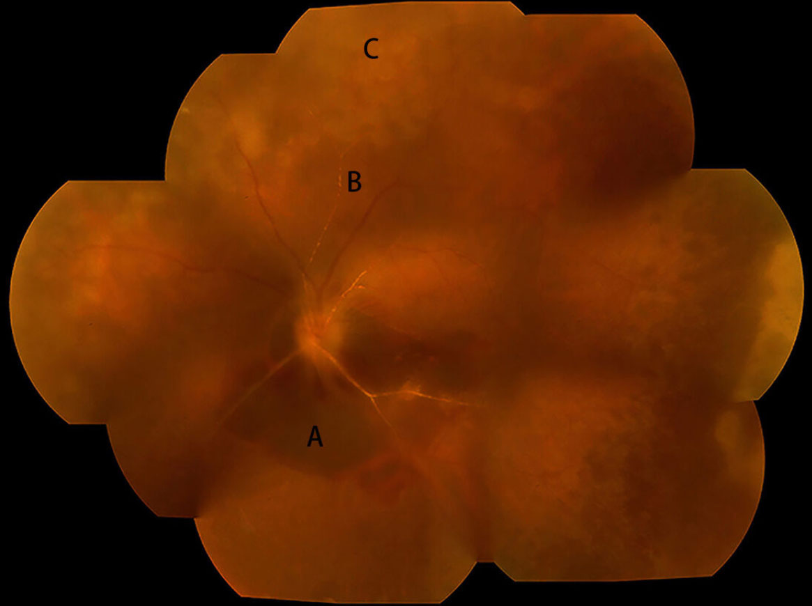
Fundus photography of ARN patients retinal necrosis of > 180° of the retinal circumference in the initially affected eye upon presentation. A: Subretinal hemorrhage lesion; B: retinal arterial sheathing; C: peripheral retinal necrosis.
3.2. Comparison of Inflammatory Indices in the ARN and Control Groups
The inflammatory parameters of the ARN and control groups are shown in Table 2. A significant inflammatory response in patients was found, which was confirmed by the altered levels of inflammatory indices; The prealbumin (P=0.019) and NC (P=0.025) of male patients were significantly higher than in female patients, whereas other baseline data showed no significant differences between male and female patients (P>0.05). There were significant differences in the NC (P=0.001), NLR (P=0.017), and GLR (P<0.001), with higher levels in the ARN group. There were no significant differences in the LC (P=0.429), MPV (P=0.627), and NAR (P=0.398) between both groups.
| Variable | Patient Group (N=20) | Control Group (N=20) | P | T or Z-value |
|---|---|---|---|---|
| Albumin (g/L) | 43.89±4.68 | 45.66±2.64 | 0.151 | -1.472 |
| Prealbumin (g/L) | 0.29±0.06 | 0.29±0.06 | 0.891 | -0.137 |
| GGT (U/L) | 23.50(19.00-32.75) | 25.00(14.75-33.50) | 0.862 | -0.176 |
| NEU count (×109L-1) | 4.56(3.28-5.62) | 3.21(2.38-3.97) | 0.001 | -3.246 |
| LYM count (×109L-1) | 2.09(1.46-2.69) | 1.85(1.59-2.21) | 0.429 | -0.798 |
| NLR median (IQR) | 2.15(1.51-3.22) | 1.79(1.14-1.97) | 0.017 | -2.38 |
| MPV (fl) | 10.61±1.01 | 10.74±0.71 | 0.627 | -0.49 |
| PLR median (IQR) | 5.45±1.75 | 5.84±1.31 | 0.429 | -0.799 |
| NAR median (IQR) | 0.10(0.08-0.12) | 0.07(0.05-0.09) | 0.398 | -3.598 |
| GLR median (IQR) | 11.13(8.39-16.90) | 12.82(9.30-17.59) | <0.001 | -0.568 |
4. DISCUSSION
ARN is a rare but devastating and potentially blinding syndrome that often occurs in immunocompetent or immunosuppressed hosts of both sexes at any age. Studies have implicated VZV, HSV, and EBV in the pathogenesis of ARN [8-13]. VZV is the principal cause of ARN in our study. The occurrence of RD upon presentation was approximately 30%. Retinal necrosis of > 180° of the retinal circumference in the initially affected eye was present in 13 of the 20 eyes (66.7%) at presentation. The ARN group had significantly higher levels of NC (p=0.001), NLR (p=0.017), and GLR (p<0.001) than those in the control group.
Our study demonstrated that VZV was the primary cause of ARN (75%), followed by HSV (5%), which is in accordance with many previous studies [8, 10, 14, 15]. Researchers conducted a national population-based study to assess the incidence of ARN in the United Kingdom and determined that VZV was the most common cause, identified in 10 patients (56%) [5].
NLR is a new and widely accepted inflammatory biomarker associated with the severity and prognosis of many cardiovascular and oncological diseases [16-19]. Knowingly, the value of the NLR has been shown with respect to retinal and ocular surface diseases, including diabetic retinopathy, age-related macular degeneration, retinal vein occlusion, glaucoma, dry eye disease, ischemic optic neuropathy, and keratoconus [20-23]. Our results showed that NLR values were significantly higher in patients with ARN than in controls, demonstrating that the occurrence of ARN is closely related to inflammation. ARN is characterized by fusion, peripheral and necrotic retinitis, peripheral occlusive arteritis, and vitreous- and anterior-chamber inflammation. However, the pathogenesis of the widespread inflammation in patients with ARN remains unclear. De Visser et al. reported that many proinflammatory and vascular mediators, such as IL-6, IL-8, IL-18, IP-10, IL-15, and ICAM-1, were significantly elevated compared to all controls [24], which predicts that proinflammatory cytokines and chemokines may be vital in the pathogenesis of ARN. Zheng and Atherton reported that CD4 + T cells, macrophages, polymorphonuclear cells, B cells, tumor necrosis factor alpha (TNF-α), and interferon-gamma (INF-γ) aid in ARN tissue necrosis [25]. Studies reported that neutrophils infiltrate and release reactive oxygen species (ROS), matrix metalloproteinases (MMPs), and neutrophil extracellular traps (NETs), leading to retinal necrosis and occlusive vasculitis [9]. Lymphocytes (especially T cells) are responsible for eliminating viruses. A decrease in lymphocytes indicates a failure in immune surveillance, making it impossible to control viral replication. Therefore, we speculated that the increase in NC and NLR is clearly correlated with the occurrence and development of ARN [26].
As a primary transferase and signal of oxidative stress, GGT has been widely reported in the prognosis of various cancers and metabolic disorders [27]. Many studies have shown that GGT can coordinate the transfer of the gamma-glutamyl moiety of the glutathione S-compound to mercapturic acid, resulting in the release of cysteinylglycine, which could cause oxidative stress and inflammation [28]. Lymphocyte count is a reliable biomarker reflecting the systemic inflammatory response in many diseases, including retinitis [29], and aids in cytotoxic cell apoptosis by inhibiting the production of inflammatory cytokines [30]. GGT is a crucial enzyme in glutathione metabolism, and its increase is a reliable indicator of systemic oxidative stress. Intracellular oxidative stress can directly trigger reactivation of the virus from the latent state by activating the virus's immediate-early genes. At the same time, oxidative stress can inhibit or even induce apoptosis of lymphocytes. The increase in GLR precisely reflects this internal environment that is beneficial for virus replication but harmful to immune defense [9]. However, GLR has not yet been reported in ophthalmic diseases. In our study, GLR in the ARN group was significantly higher than that in the control group, indicating that the occurrence and progression of ARN are associated with inflammation.
This study has several limitations. First, the retrospective, single-center design introduces inherent biases that limit the generalizability of our results. Second, selection bias is a significant concern. This hospital-based sampling potentially inflated effect sizes, limiting the applicability of our results to the general population. Third, the absence of longitudinal follow-up data restricts our ability to assess dynamic changes in inflammatory biomarkers over time. Fourth, while we identified significant associations with inflammatory markers, we were unable to establish validated clinical cutoff values for risk stratification. Fifth, we acknowledge the presence of unmeasured confounding variables like medication history that may have influenced our results.
CONCLUSION
This article focuses on inflammatory parameters in the blood of patients with ARN, is a serious ocular disease that causes visual impairment, so understanding and appropriate treatment are pivotal. In our study, we found that NLR and GLR, with significant differences between the two groups, are closely associated with ARN. These two blood parameters may be indicators for the diagnosis of ARN, emphasizing the importance of early blood examination.
AUTHORS’ CONTRIBUTIONS
The authors confirm their contribution to the paper as follows: L.C., W.C.: Significant contribution to conception and design; T.Z.: Data acquisition; T.Z., W.H.: Data analysis and interpretation; T.Z., W.H.: Drafting; L.C., W.C.: Significant intellectual content revision of the manuscript; L.C., W.C., T.Z., W.H.: Final approval of the submitted manuscript; T.Z.: Statistical analysis; L.C., W.C.: Obtaining funding; L.C., W.C.: Supervision of administrative, technical, or material support; L.C., W.C.: Research group leadership. All authors reviewed the results and approved the final version of the manuscript.
LIST OF ABBREVIATIONS
| ARN | = Acute Retinal Necrosis |
| BCVA | = Best-Corrected Visual Acuity |
| CMV | = Cytomegalovirus |
| DNA | = Deoxyribonucleic Acid |
| EBV | = Epstein-Barr Virus |
| GGT | = Gamma-glutamyl transferase |
| GLR | = Gamma-glutamyl transferase to Lymphocyte Count Ratio |
| HSV | = Herpes Simplex Virus |
| ICAM-1 | = Intercellular Adhesion Molecule-1 |
| IL-6 | = Interleukin-6 |
| IL-8 | = Interleukin-8 |
| IL-15 | = Interleukin-15 |
| IL-18 | = Interleukin-18 |
| INF-γ | = Interferon Gamma |
| IOP | = Intraocular Pressure |
| IP-10 | = Interferon Gamma-Inducible Protein 10 |
| LC | = Lymphocyte Count |
| MMPs | = Matrix Metalloproteinases |
| MPV | = Mean Platelet Volume |
| NAR | = Neutrophil-to-Albumin Ratio |
| NC | = Neutrophil Count |
| NETs | = Neutrophil Extracellular Traps |
| NLR | = Neutrophil-to-Lymphocyte Ratio |
| PCR | = Polymerase Chain Reaction |
| RD | = Retinal Detachment |
| ROS | = Reactive Oxygen Species |
| SPSS | = Statistical Package for the Social Sciences |
| TNF-α | = Tumor Necrosis Factor Alpha |
| VZV | = Varicella-Zoster Virus |
ETHICS APPROVAL AND CONSENT TO PARTICIPATE
This study was approved by the Ethics Committee of Beijing Chaoyang Hospital, Capital Medical University, China (Approval No. 2018-KE-125).
HUMAN AND ANIMAL RIGHTS
All human research procedures followed were in accordance with the ethical standards of the committee responsible for human experimentation (institutional and national), and with the Helsinki Declaration of 1975, as revised in 2013.
AVAILABILITY OF DATA AND MATERIALS
The data supporting the findings of the article is available in the [Zenodo Repository] at [https://zenodo.org/records/21862682], reference number [DOI: 10.5281/zenodo.21862682].
ACKNOWLEDGEMENTS
Declared none.